Copyright
©2011 Baishideng Publishing Group Co.
World J Gastroenterol. Jan 28, 2011; 17(4): 499-505
Published online Jan 28, 2011. doi: 10.3748/wjg.v17.i4.499
Published online Jan 28, 2011. doi: 10.3748/wjg.v17.i4.499
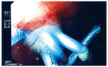
Figure 1 Pus drained from an infected pseudocyst.
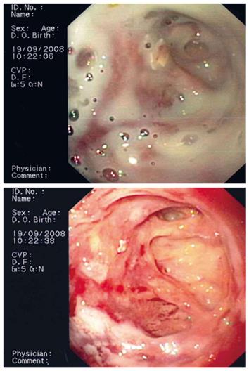
Figure 2 An infected pseudocyst before and after flushing.
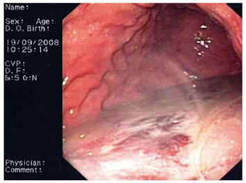
Figure 3 Pus accumulating in the stomach.
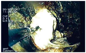
Figure 4 Necrosectomy in a pseudocyst.
- Citation: Sadik R, Kalaitzakis E, Thune A, Hansen J, Jönson C. EUS-guided drainage is more successful in pancreatic pseudocysts compared with abscesses. World J Gastroenterol 2011; 17(4): 499-505
- URL: https://www.wjgnet.com/1007-9327/full/v17/i4/499.htm
- DOI: https://dx.doi.org/10.3748/wjg.v17.i4.499


