Copyright
©2011 Baishideng Publishing Group Co.
World J Gastroenterol. Apr 21, 2011; 17(15): 2019-2027
Published online Apr 21, 2011. doi: 10.3748/wjg.v17.i15.2019
Published online Apr 21, 2011. doi: 10.3748/wjg.v17.i15.2019
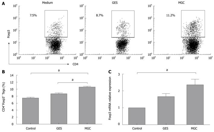
Figure 1 Gastric cancer cell supernatant induces the increased Tregs in coculture with peripheral blood mononuclear cells.
A: Representative flow cytometry analysis of CD4+Foxp3+ Tregs frequency in CD4+ T cells population following peripheral blood mononuclear cells co-culture with medium, GES, or MGC supernatants. Rectangles show double positive gating and numbers reflect percentage of cells in that gate; B: Summarized data from all subjects showed that CD4+Foxp3+ Tregs increased in coculture with MGC supernatants (aP < 0.05); C: Relative quantity of Foxp3 mRNA was measured by real-time polymerase chain reaction before and after culture with medium, GES, or MGC supernatants.
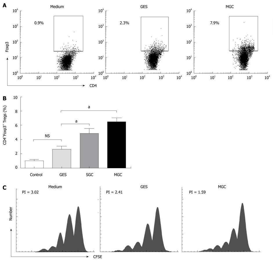
Figure 2 Gastric cancer cell supernatant mediates the conversion of CD4+CD25- T cell to CD4+Foxp3+ Tregs.
A: Representative flow cytometry analysis of CD4+Foxp3+ Tregs frequency in CD4+ T cells population following sorted CD4+CD25-CD127+ T cells co-culture with medium, GES, or MGC supernatants; B: Summarized data showed that both MGC and SGC supernatants induced higher CD4+Foxp3+ Tregs (aP < 0.05); C: After co-culture with MGC, GES or medium, CD4+CD25- T cells were placed in coculture with CFSE-labeled CD4+CD25-CD127+ T cells at a ratio of 1:1 in the presence of soluble anti-CD3/CD28 as well as IL-2. The representative data from three independent experiments are shown.
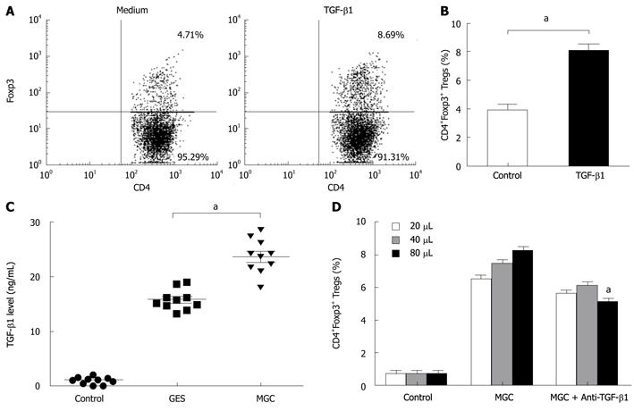
Figure 3 Gastric cancer cells producing transforming growth factor-β1 can partially mediate the conversion of CD4+CD25- T cells to CD4+Foxp3+ Treg cells.
A: Representative flow cytometry analysis of CD4+Foxp3+ Tregs frequency in the presence or absence of transforming growth factor-β1 (TGF-β1); B: Summarized data showed that CD4+Foxp3+ Tregs frequency increased in presence of TGF-β1 in comparison with that in absence of TGF-β1 (aP < 0.05); C: Gastric cancer cell TGF-β1 production was assessed by enzyme-linked immunosorbent assay in supernatants. (aP < 0.05); D: TGF-β1 blocking antibody or control IgG1 antibody was added to the co-culture in order to monitor impairment of Tregs development within the coculture system. Different volumes of supernatant protein concentrate (20, 40, and 80 μL) from MGC were added to the co-culture system. (aP < 0.05).
- Citation: Yuan XL, Chen L, Zhang TT, Ma YH, Zhou YL, Zhao Y, Wang WW, Dong P, Yu L, Zhang YY, Shen LS. Gastric cancer cells induce human CD4+Foxp3+ regulatory T cells through the production of TGF-β1. World J Gastroenterol 2011; 17(15): 2019-2027
- URL: https://www.wjgnet.com/1007-9327/full/v17/i15/2019.htm
- DOI: https://dx.doi.org/10.3748/wjg.v17.i15.2019


