Copyright
©2011 Baishideng Publishing Group Co.
World J Gastroenterol. Mar 14, 2011; 17(10): 1379-1382
Published online Mar 14, 2011. doi: 10.3748/wjg.v17.i10.1379
Published online Mar 14, 2011. doi: 10.3748/wjg.v17.i10.1379
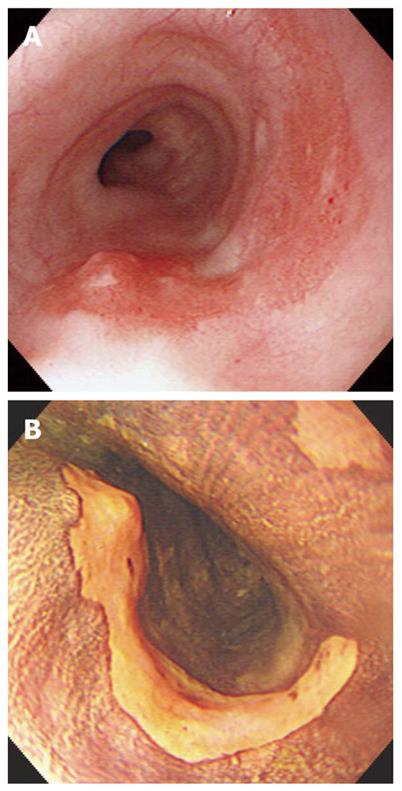
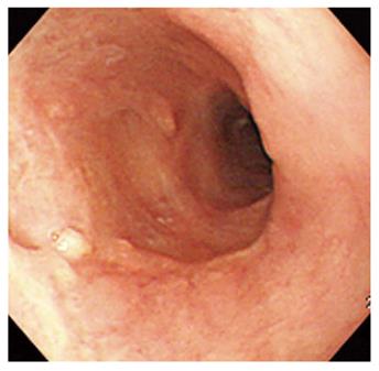
Figure 1 Gastroscopy before photodynamic therapy.
A: Gastroscopy showed a flat and reddish lesion in the mid-esophagus (a biopsy showed squamous cell carcinoma); B: Gastroscopy showed an unstained lesion after spraying Lugol solution in the mid-esophagus.
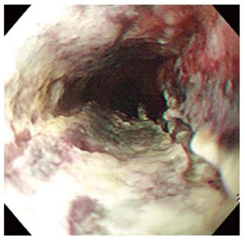
Figure 2 Two days after Photodynamic therapy.
Endoscopy showed circumferential coagulation necrosis with an ulcer in the photodynamic therapy-treated lesion.
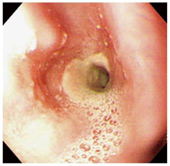
Figure 3 Two months after Photodynamic therapy.
Endoscopy showed luminal narrowing with fibrous scarring changes in the photodynamic therapy-treated lesion.
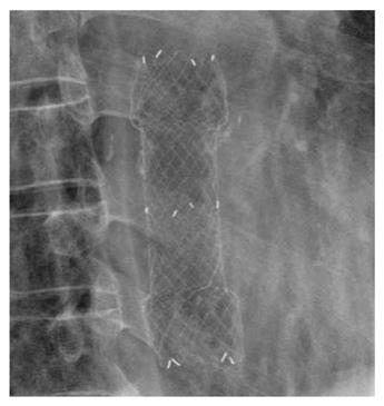
Figure 4 Fluoroscopic image showed a metal stent at the stricture site in the esophagus.
Figure 5 Endoscopy showed improvement of the previous stricture site 2 mo after stent removal.
- Citation: Cheon YK. Metal stenting to resolve post-photodynamic therapy stricture in early esophageal cancer. World J Gastroenterol 2011; 17(10): 1379-1382
- URL: https://www.wjgnet.com/1007-9327/full/v17/i10/1379.htm
- DOI: https://dx.doi.org/10.3748/wjg.v17.i10.1379


