Copyright
©2009 The WJG Press and Baishideng.
World J Gastroenterol. Nov 21, 2009; 15(43): 5425-5431
Published online Nov 21, 2009. doi: 10.3748/wjg.15.5425
Published online Nov 21, 2009. doi: 10.3748/wjg.15.5425
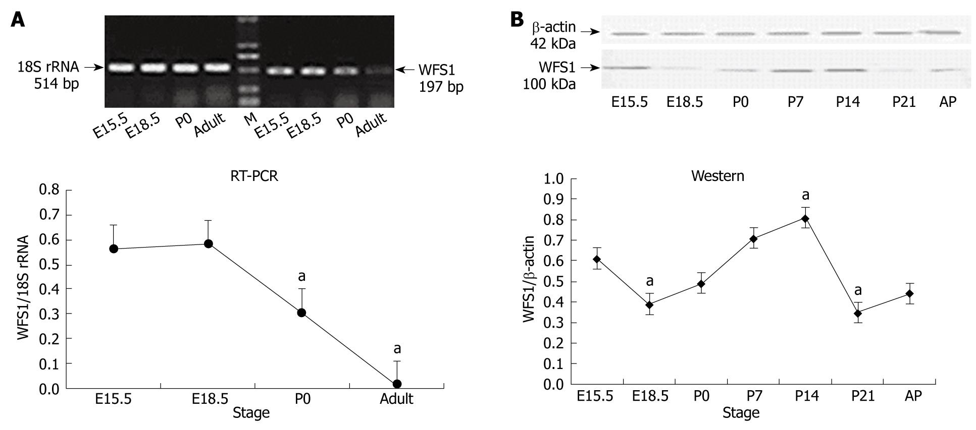
Figure 1 Wolfram syndrome 1 (WFS1) mRNA (A) and protein expression (B) in the developing rat pancreas.
A: WFS1 mRNA measured by reverse transcription-polymerase chain reation (RT-PCR). upper: from embryonic day (E) 15.5 to adulthood: WFS1 mRNA expression was highest at E15.5 and E18.5. M: Marker; lower: WFS1 mRNA expression was analyzed and normalized to 18S rRNA. Results are indicated in percentage above the 18sRNA value and are representative of three independent experiments. Compared to E15.5, the highest level of WFS1 mRNA was detected at E18.5, the E15.5 and P0 was less, and at a lowest at adult (aP < 0.05 vs P0 and adult), respectively; B: Western blotting of WFS1 protein in the pancreas of E15.5, E18.5, P0, P14, P21 and adult rats. upper: expression of WFS1 protein (100 kDa) with β-actin (42 kDa) as a loading control. The position of the molecular weight markers are indicated on the left; lower: the expression of WFS1 protein shows dynamic changes during rat pancreas development. Results are indicated in percentage above the e15.5 value and are representative of three independent experiments. Compare to E15.5, The highest level of WFS1 was at P14 and lowest at P21 (aP < 0.05 vs E18.5, P14 and P21), respectively.
Figure 2 Immunofluorescent localization of WFS1 protein and insulin in the pancreas of E18.
5, P0, P14, P21 and adult rats. The WFS1 antibody was detected with an FITC (green)-labeled secondary antibody and the insulin antibody was detected with a Cy3 (red)-labeled secondary antibody. An overlap between WFS1 (green) and insulin (red) labeling is colored orange. Modest WFS1 staining is seen in the developing islets from E18.5 to adulthood, and overlaps insulin-positive β-cells. A few WFS1-labeled cells are scattered away from the islets. Primary objective magnification, × 400. Insets, higher magnifications of the areas indicated by arrows for E18.5, P0 and P14.
Figure 3 Immunofluorescent localization of WFS1 protein and glucagon in the pancreas of P0 and adult rats.
The WFS1 antibody was detected with an FITC (green)-labeled secondary antibody and the glucagon antibody (mouse monoclonal antibody) was detected with a Cy3 (red)-labeled secondary antibody on the same section. The overlap between WFS1 (green) and glucagon (red) labeling is colored orange. WFS1-positive cells and glucagon-positive cells can be seen in 2 pancreas sections tested. Double-labeling with antibodies to WFS1 and glucagon reveal that the cells labeled with WFS1 are surrounded by α-cells. Primary objective magnification, × 400.
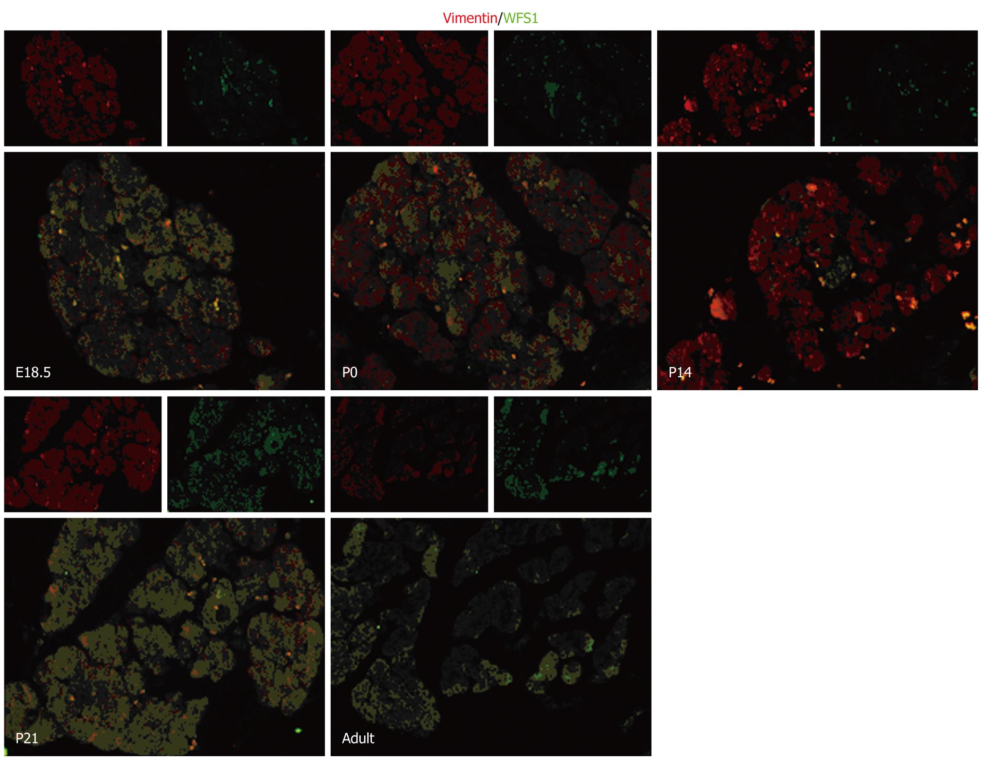
Figure 4 Immunofluorescent localization of WFS1 protein and vimentin in the pancreas of E18.
5, P0, P14, P21 and adult rats. The WFS1 antibody was detected with an FITC (green)-labeled secondary antibody and the vimentin antibody was detected with a Cy3 (red)-labeled secondary antibody on the same section. The overlap between AFP (green) and vimentin (red) labeling is colored orange. WFS1-positive cells can be detected in pancreas sections from E18.5 to adult. Double-labeling with antibodies to WFS1 and to vimentin revealed colocalization of these proteins in the same cells from E18.5 to adult. Primary objective magnification, × 400.
-
Citation: Xu R, Xia B, Geng J, Shi J, Shi H, Yuan L, De W. Expression and localization of
Wolfram syndrome 1 gene in the developing rat pancreas. World J Gastroenterol 2009; 15(43): 5425-5431 - URL: https://www.wjgnet.com/1007-9327/full/v15/i43/5425.htm
- DOI: https://dx.doi.org/10.3748/wjg.15.5425


