Copyright
©2008 The WJG Press and Baishideng.
World J Gastroenterol. Jan 28, 2008; 14(4): 518-523
Published online Jan 28, 2008. doi: 10.3748/wjg.14.518
Published online Jan 28, 2008. doi: 10.3748/wjg.14.518
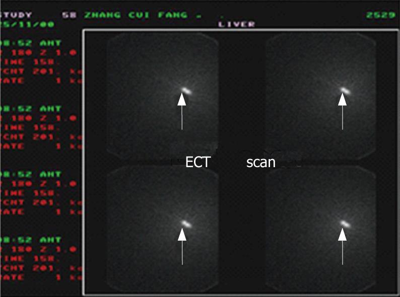
Figure 1 Liver ECT scan.
Arrow: The site where P32-GMS was implanted.
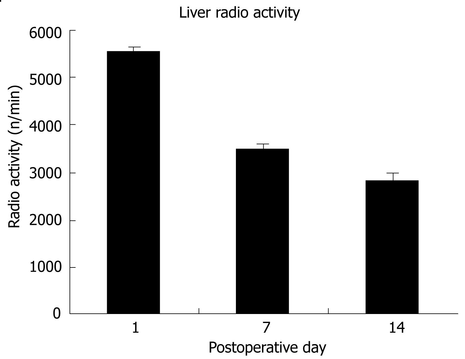
Figure 2 P32 Radioactivity in liver of the patients with P32-GMS (n/min).
The radio activity of P32-GMS decreased on POD 7 and POD 14 as compared with POD 1. (POD = postoperative day).
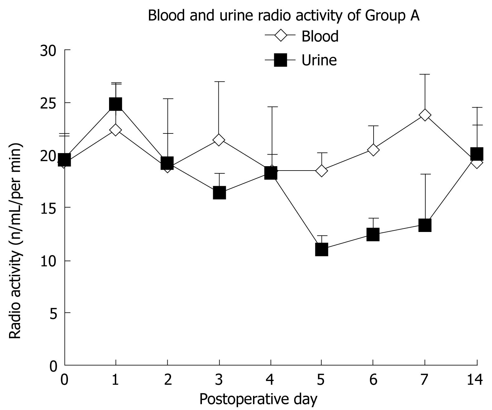
Figure 3 Radioactivity in blood and urine of the patients with P32-GMS (n/mL/min).
The radio activity in blood and urine were low. It was familiar with the sample without P32-GMS implanted (d 0).
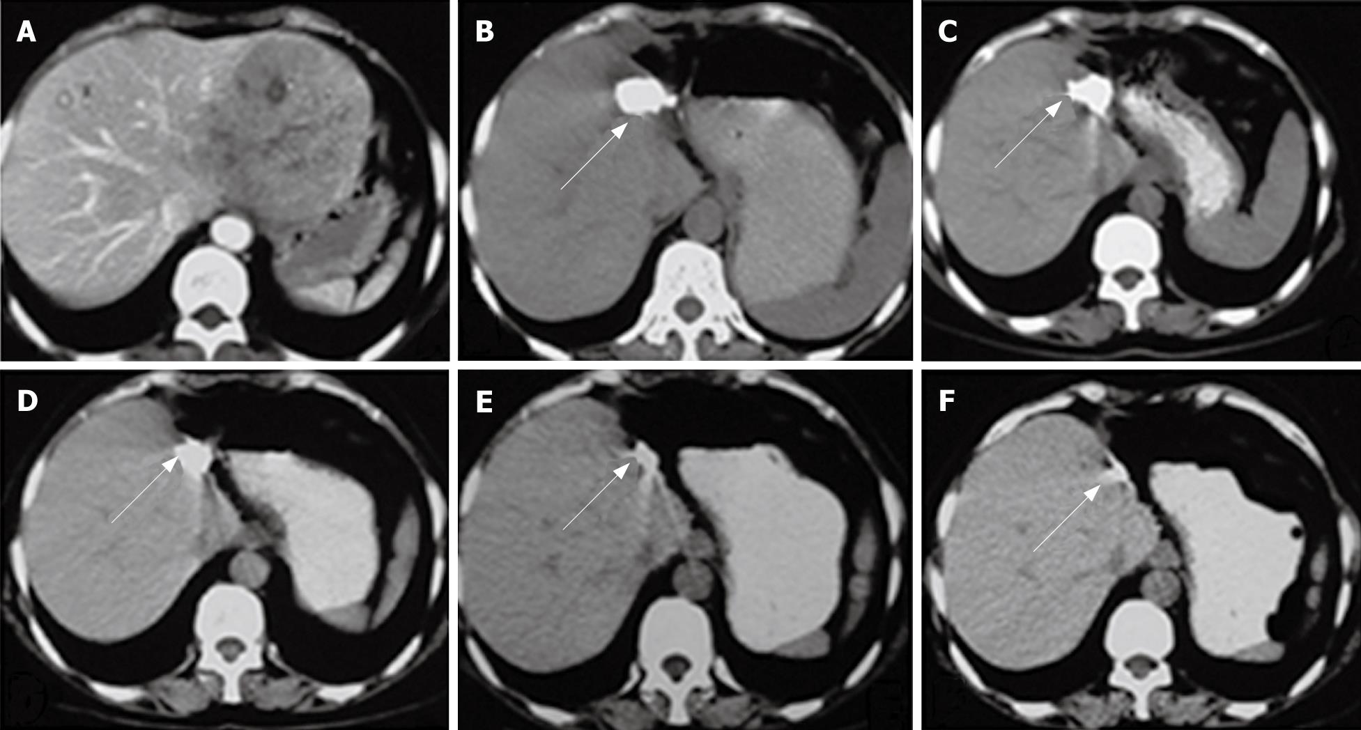
Figure 4 CT scan of the HCC patient who underwent hepatectomy and P32-GMS treatments in 4 years following-up.
A: Liver CT scan showing a progressive liver tumor prior to hepatectomy; B: Liver CT scan after hepatectomy; C: Liver CT scan at first postoperative year; D: Liver CT at second postoperative year; E: Liver CT at third postoperative year; F: Liver CT at fourth postoperative year. Arrows: implanted P32-GMS.
- Citation: Wang XM, Yin ZY, Yu RX, Peng YY, Liu PG, Wu GY. Preventive effect of regional radiotherapy with phosphorus-32 glass microspheres in hepatocellular carcinoma recurrence after hepatectomy. World J Gastroenterol 2008; 14(4): 518-523
- URL: https://www.wjgnet.com/1007-9327/full/v14/i4/518.htm
- DOI: https://dx.doi.org/10.3748/wjg.14.518


