Copyright
©2008 The WJG Press and Baishideng.
World J Gastroenterol. Jul 14, 2008; 14(26): 4234-4237
Published online Jul 14, 2008. doi: 10.3748/wjg.14.4234
Published online Jul 14, 2008. doi: 10.3748/wjg.14.4234
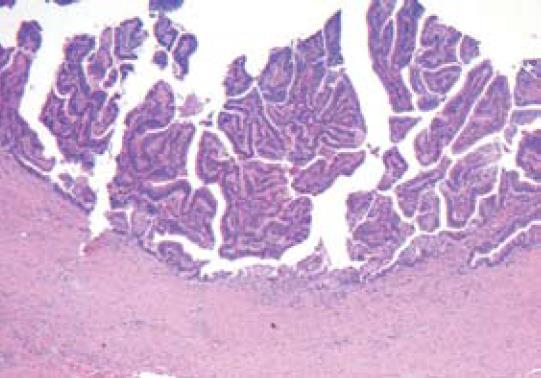
Figure 1 HE staining for samples collected at the right-sided hemihepatectomy and hepatico-jejunostomy of the left hepatic duct (× 40) showing a villous adenoma with mild inflammation of the adjacent proximal parts of the bile duct and the surrounding liver tissue but no invasive growth.
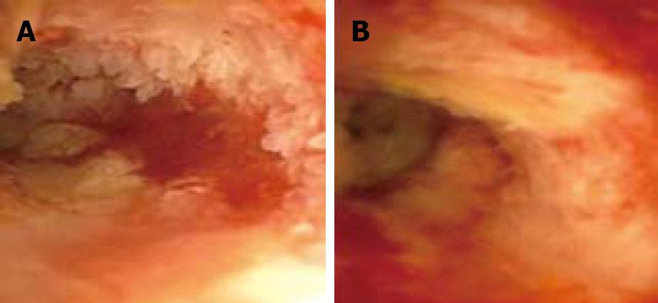
Figure 2 Recurrent papillomatosis before the third course of photodynamic therapy without signs of a functional stenosis of the bile duct (A) and one month after the second course of PDT (B).
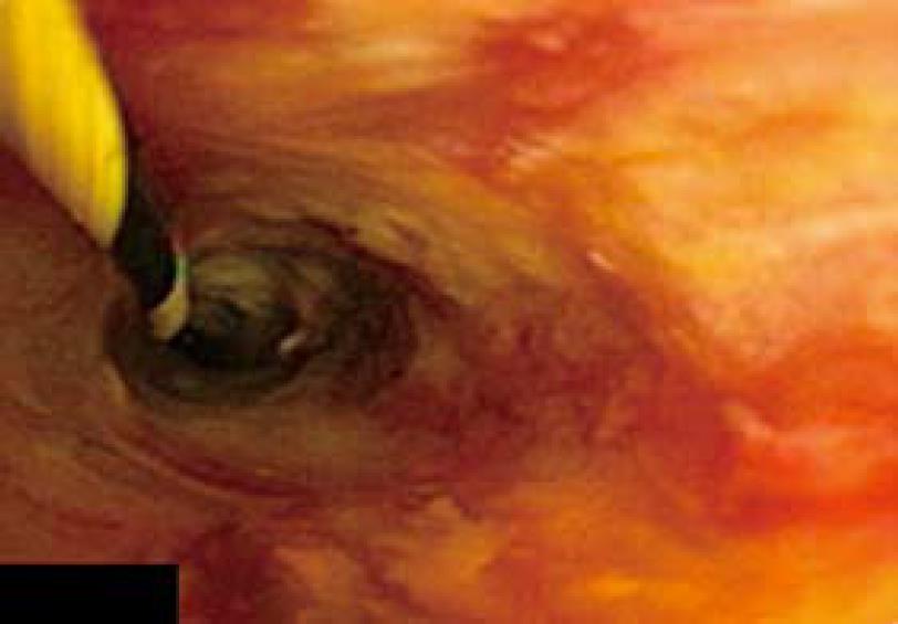
Figure 3 Cholangioscopy showing only minor adenomatosis 3 mo after the third course of PDT.
- Citation: Bechmann LP, Hilgard P, Frilling A, Schumacher B, Baba HA, Gerken G, Zoepf T. Successful photodynamic therapy for biliary papillomatosis: A case report. World J Gastroenterol 2008; 14(26): 4234-4237
- URL: https://www.wjgnet.com/1007-9327/full/v14/i26/4234.htm
- DOI: https://dx.doi.org/10.3748/wjg.14.4234


