Copyright
©2008 The WJG Press and Baishideng.
World J Gastroenterol. Jan 7, 2008; 14(1): 125-128
Published online Jan 7, 2008. doi: 10.3748/wjg.14.125
Published online Jan 7, 2008. doi: 10.3748/wjg.14.125
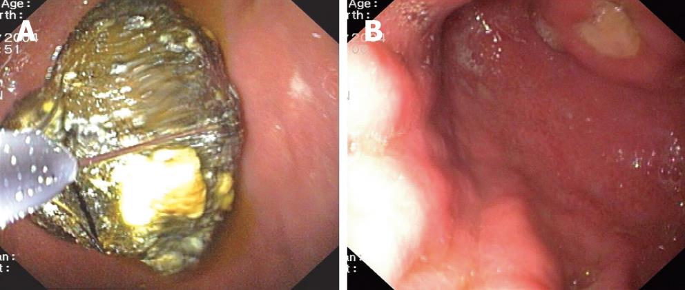
Figure 1 Oesophagogastroduodenoscopy confirming the presence of a foreign body looking like a biliary stone (A) and a 15 mm-wide ulcer (B).
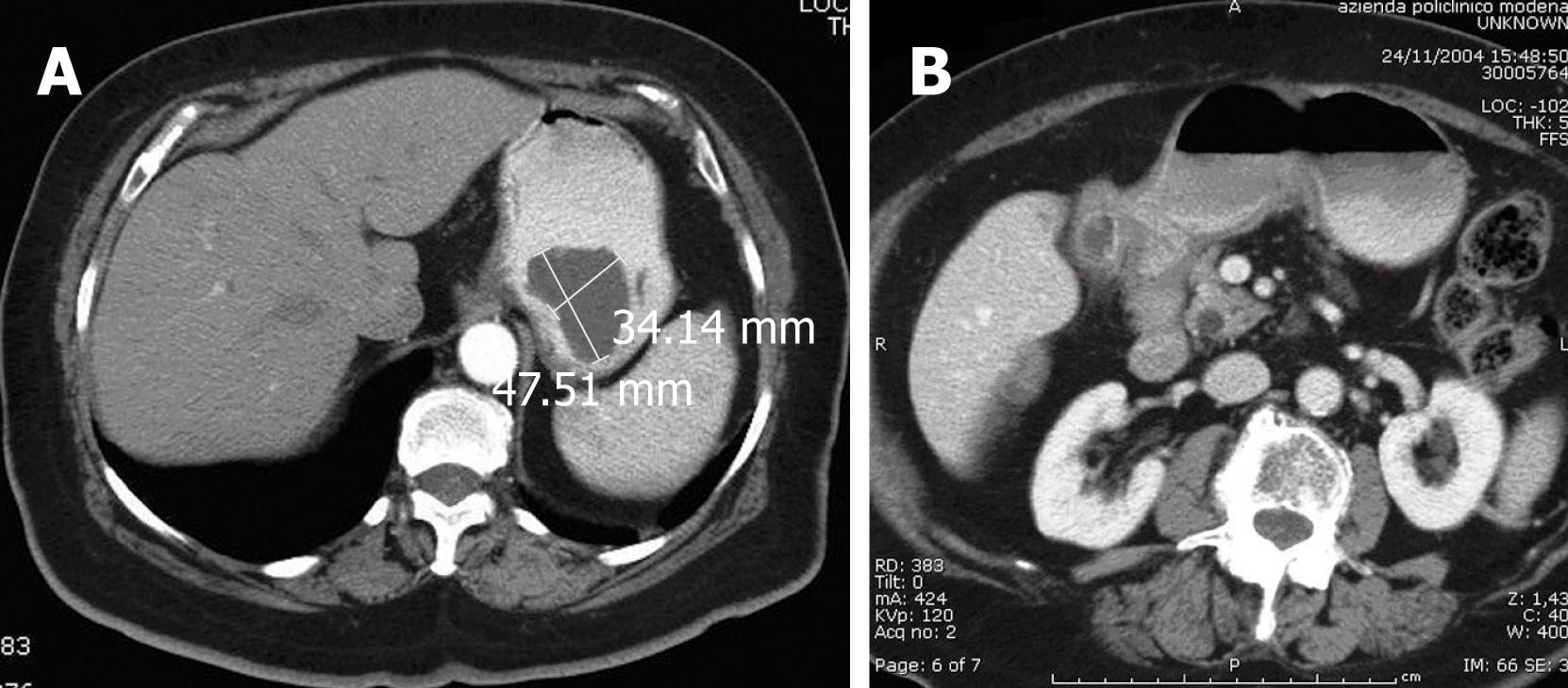
Figure 2 Computerised tomography revealing the density of foreign body typical of ectopic biliary stone (A) and the oedematous wall of the gastric antrum (B).
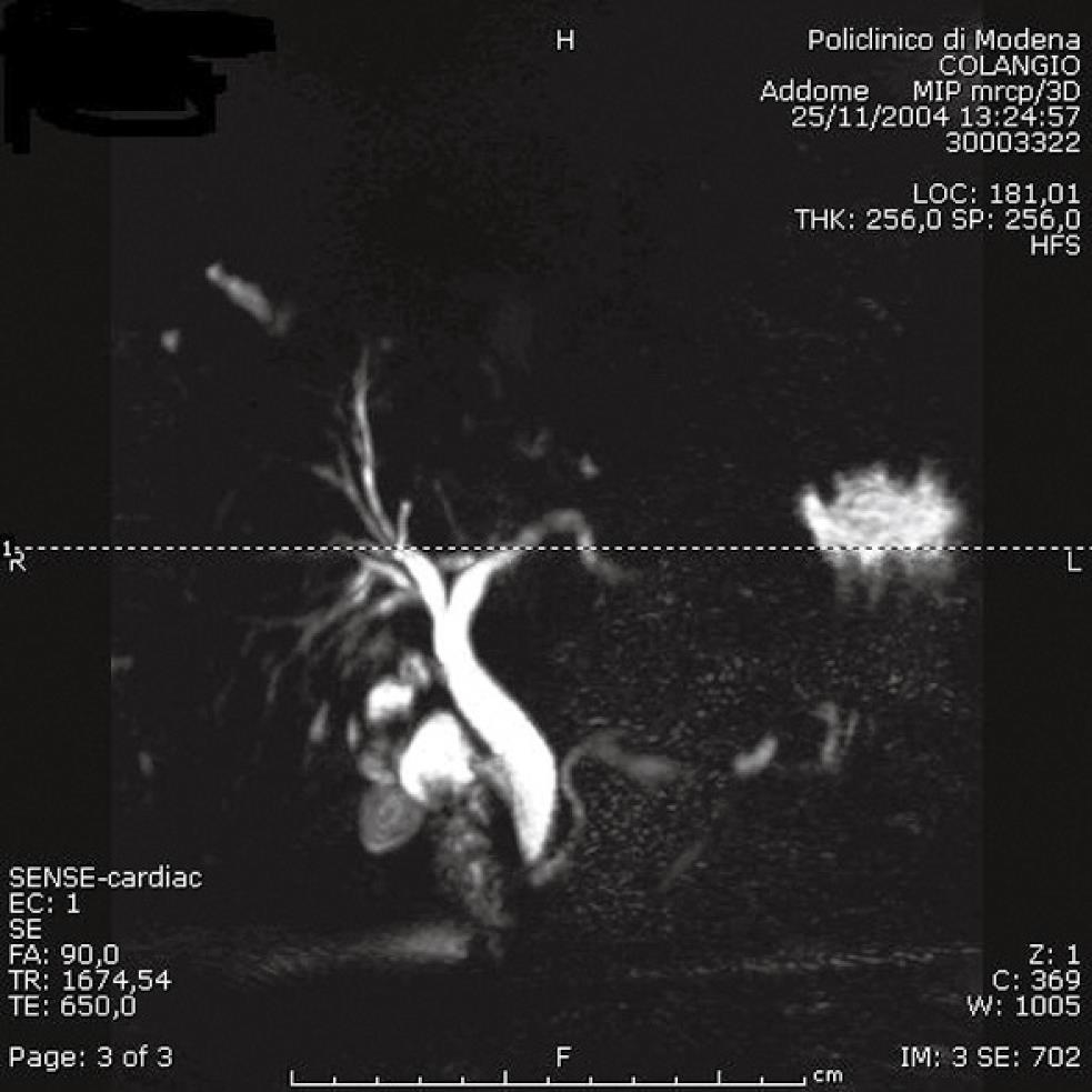
Figure 3 Magnetic resonance cholangiopancreatography showing no fistula.
Figure 4 One-stage surgery with dissection of omental-cholecystic adhesions.
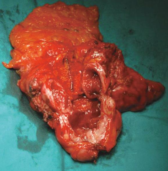
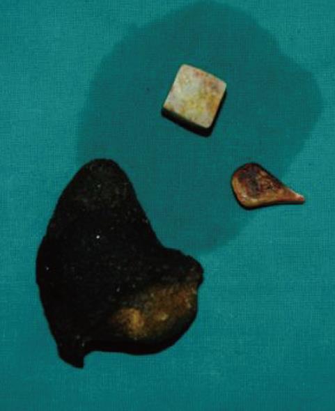
Figure 5 A large ectopic stone extracted.
- Citation: Arioli D, Venturini I, Masetti M, Romagnoli E, Scarcelli A, Ballesini P, Borghi A, Barberini A, Spina V, Santis MD, Benedetto FD, Gerunda GE, Zeneroli ML. Intermittent gastric outlet obstruction due to a gallstone migrated through a cholecysto-gastric fistula: A new variant of “Bouveret’s syndrome”. World J Gastroenterol 2008; 14(1): 125-128
- URL: https://www.wjgnet.com/1007-9327/full/v14/i1/125.htm
- DOI: https://dx.doi.org/10.3748/wjg.14.125


