Copyright
©2007 Baishideng Publishing Group Co.
World J Gastroenterol. Mar 7, 2007; 13(9): 1460-1465
Published online Mar 7, 2007. doi: 10.3748/wjg.v13.i9.1460
Published online Mar 7, 2007. doi: 10.3748/wjg.v13.i9.1460
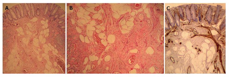
Figure 1 Histopathologic appearance of small rectal pedunculated angiolipoma (macroscopically 7 mm x 5 mm x 4 mm in diameter with normal mucosal appearence).
A: Proliferated blood vessels and adipose cells in mature adipose tissue in submucosa of colon at small magnification (light microscope, HE); B: Proliferated vascular tissues and mature adipose tissue in submucosa. There was no fibrin trombus in blood vessels (light microscope, HE); C: SMA staining of angiolipoma.
- Citation: Kacar S, Kuran S, Temucin T, Odemis B, Karadeniz N, Sasmaz N. Rectal angiolipoma: A case report and review of literature. World J Gastroenterol 2007; 13(9): 1460-1465
- URL: https://www.wjgnet.com/1007-9327/full/v13/i9/1460.htm
- DOI: https://dx.doi.org/10.3748/wjg.v13.i9.1460


