Copyright
©2007 Baishideng Publishing Group Inc.
World J Gastroenterol. Jul 14, 2007; 13(26): 3645-3648
Published online Jul 14, 2007. doi: 10.3748/wjg.v13.i26.3645
Published online Jul 14, 2007. doi: 10.3748/wjg.v13.i26.3645
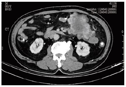
Figure 1 Abdominal CT scan showed a 11 cm heterogeneous mass at the mesentery without bowel involvement.
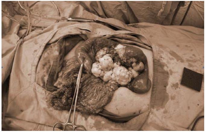
Figure 2 A multinodular tumor at the mesentery of proximal jejunum, 11 cm × 11 cm × 7 cm in size, with irregular margin was found on laparotomy, the omentum was focally involved.
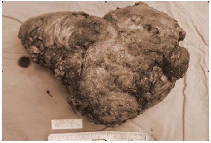
Figure 3 The surgical specimen showed a tumor 19 cm × 17 cm × 10 cm in size involving the mesentery of both jejunum and ileum.
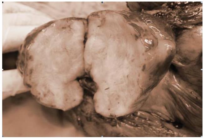
Figure 4 The saggital section showed that the tumor was well circumscribed, grayish white, and firm with foci of hemorrhage, necrosis, and cystic degeneration.
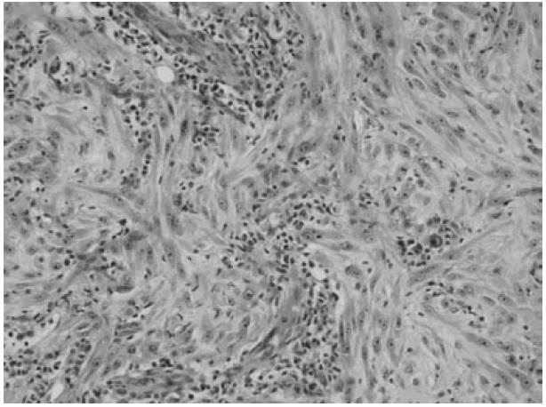
Figure 5 Microscopically, the spindled myofibroblasts and ganglion-like cells dispersed in a myxoid background with inflammatory reaction.
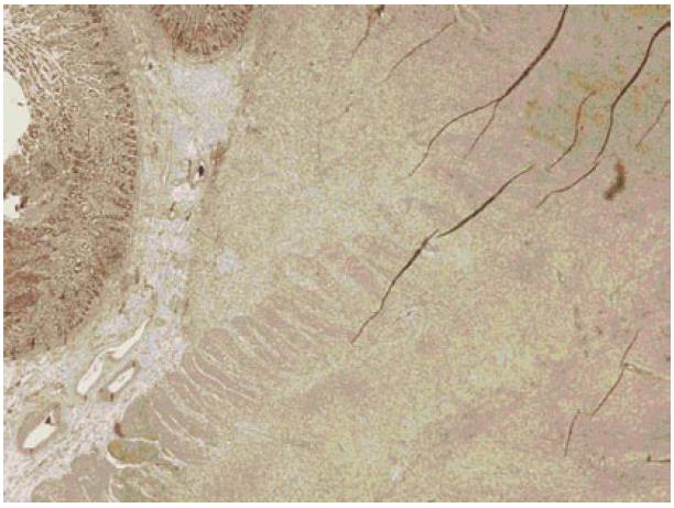
Figure 6 The spindled myofibroblasts invaded the muscle layer of bowel wall.
- Citation: Chen SS, Liu SI, Mok KT, Wang BW, Yeh MH, Chen YC, Chen IS. Mesenteric inflammatory myofibroblastic tumors in an elder patient with early recurrence: A case report. World J Gastroenterol 2007; 13(26): 3645-3648
- URL: https://www.wjgnet.com/1007-9327/full/v13/i26/3645.htm
- DOI: https://dx.doi.org/10.3748/wjg.v13.i26.3645


