Copyright
©2007 Baishideng Publishing Group Inc.
World J Gastroenterol. Jul 14, 2007; 13(26): 3638-3640
Published online Jul 14, 2007. doi: 10.3748/wjg.v13.i26.3638
Published online Jul 14, 2007. doi: 10.3748/wjg.v13.i26.3638
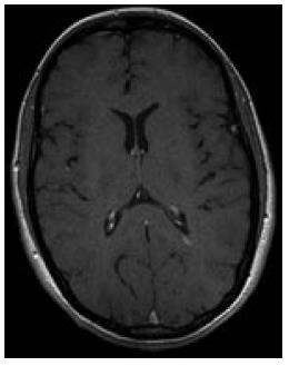
Figure 1 T1-weighted axial MRI showing contrast enhancing lesion indicative of inflammatory activity in MS.
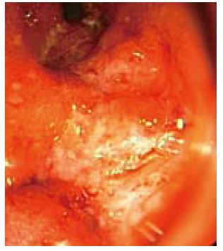
Figure 2 Severe ulcerative colitis on colonoscopy.
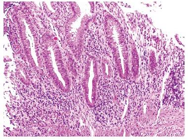
Figure 3 Presence of ulcerative colitis hallmark features on histology (HE, x 5).
- Citation: Schott E, Paul F, Wuerfel JT, Zipp F, Rudolph B, Wiedenmann B, Baumgart DC. Development of ulcerative colitis in a patient with multiple sclerosis following treatment with interferonβ 1a. World J Gastroenterol 2007; 13(26): 3638-3640
- URL: https://www.wjgnet.com/1007-9327/full/v13/i26/3638.htm
- DOI: https://dx.doi.org/10.3748/wjg.v13.i26.3638


