Copyright
©2007 Baishideng Publishing Group Co.
World J Gastroenterol. Apr 14, 2007; 13(14): 2077-2082
Published online Apr 14, 2007. doi: 10.3748/wjg.v13.i14.2077
Published online Apr 14, 2007. doi: 10.3748/wjg.v13.i14.2077
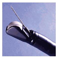
Figure 1 Toshiba-Fujinon curved linear array echoendoscope PEF-708FA.
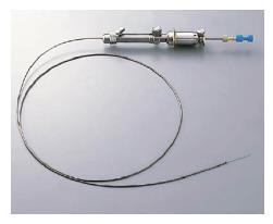
Figure 2 Olympus power shot type metallic needle NA-11J-KB.
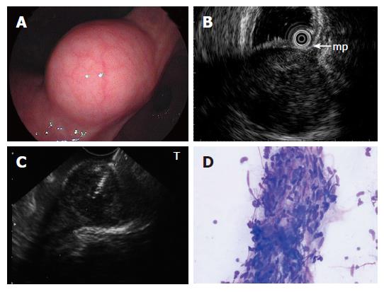
Figure 3 A: Submucosal lesion in the angulus of the stomach shown on endoscopy; B: EUS using ultrasound catheter probe reveals 3 cm subepithelial hypoechoic tumor with continuity to proper muscle layer (arrow-mp); C: Puncture of the small GIST under direct endosonographic visualization.
The needle can be visualized; D: EUS-FNA smear of GIST showing a small tissue fragment composed of ovoid to spindle-shaped nuclei with minimal to no atypia arranged in fascicles (modified Giemsa stain).
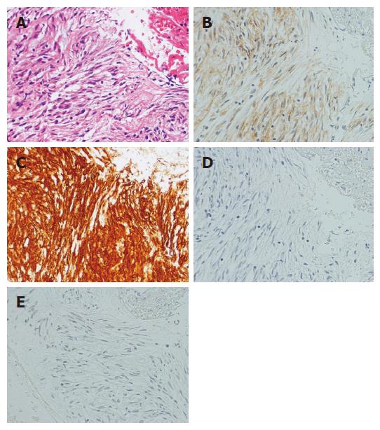
Figure 4 Photomicrographs of EUS-FNA specimen of GIST.
A: Hematoxylin-eosin stain; B: Immunohistochemical stain for c-kit; C: Immunohistochemical stain for CD34; D: Immunohistochemical stain muscle actin; E: Immunohistochemical stain for S-100. The tumor is diffusely positive for c-kit and CD34 and negative for muscle actin and S-100. The immunohistochemical pattern diagnosis is GIST.
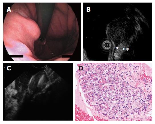
Figure 5 A: Endoscopy showing submucosal lesion in the upper body of the stomach; B: EUS using ultrasound catheter probe demonstrating 1.
5 cm subepithelial hypoechoic tumor with continuity to proper muscle layer (arrow-mp); C: The hypoechoic mass shown on EUS was punctured under real-time EUS guidance; D: The FNAB specimen consisted of acinar cells. The histologic diagnosis was ectopic pancreas.
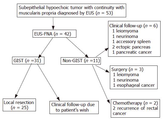
Figure 6 EUS-FNA diagnosis and following management.
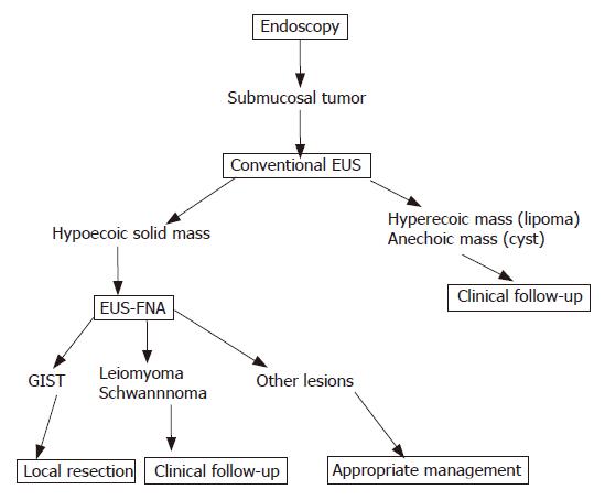
Figure 7 Algorithm of diagnosis and treatment of gastrointestinal submucosal tumor.
- Citation: Akahoshi K, Sumida Y, Matsui N, Oya M, Akinaga R, Kubokawa M, Motomura Y, Honda K, Watanabe M, Nagaie T. Preoperative diagnosis of gastrointestinal stromal tumor by endoscopic ultrasound-guided fine needle aspiration. World J Gastroenterol 2007; 13(14): 2077-2082
- URL: https://www.wjgnet.com/1007-9327/full/v13/i14/2077.htm
- DOI: https://dx.doi.org/10.3748/wjg.v13.i14.2077


