Copyright
©2006 Baishideng Publishing Group Co.
World J Gastroenterol. Dec 21, 2006; 12(47): 7642-7648
Published online Dec 21, 2006. doi: 10.3748/wjg.v12.i47.7642
Published online Dec 21, 2006. doi: 10.3748/wjg.v12.i47.7642
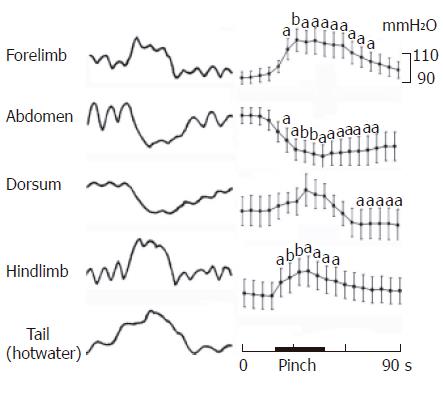
Figure 2 Gastric motility in response to cutaneous natural stimuli.
aP < 0.05, bP < 0.01 vs control gastric motility of pre-stimuli.
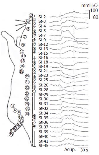
Figure 3 The gastric motility in response to craniocaudal stomach-meridian acupoints stimuli.
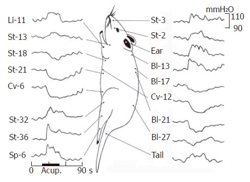
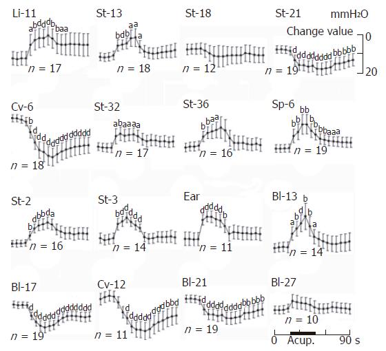
Figure 4 Gastric motility in response to acu-stimulation.
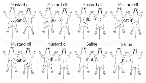
Figure 1 Schematic drawing of number and location of blue dots in rats.
Dye extravasation in skin after inflammation of intragastric mucous membrane and rare small blue dots over abdominal area of control rats.
Figure 5 The quantitative analysis of gastric motility in response to acu-stimulation.
aP < 0.05, bP < 0.01, dP < 0.001 vs control gastric motility of pre-stimuli.
- Citation: Li YQ, Zhu B, Rong PJ, Ben H, Li YH. Effective regularity in modulation on gastric motility induced by different acupoint stimulation. World J Gastroenterol 2006; 12(47): 7642-7648
- URL: https://www.wjgnet.com/1007-9327/full/v12/i47/7642.htm
- DOI: https://dx.doi.org/10.3748/wjg.v12.i47.7642


