Copyright
©2006 Baishideng Publishing Group Co.
World J Gastroenterol. Jan 28, 2006; 12(4): 662-664
Published online Jan 28, 2006. doi: 10.3748/wjg.v12.i4.662
Published online Jan 28, 2006. doi: 10.3748/wjg.v12.i4.662
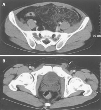
Figure 1 Contrast-enhanced computed tomography showing a large fat density mass below the sigmoid colon and left inguinal hernia with incarcerated fat (arrow).
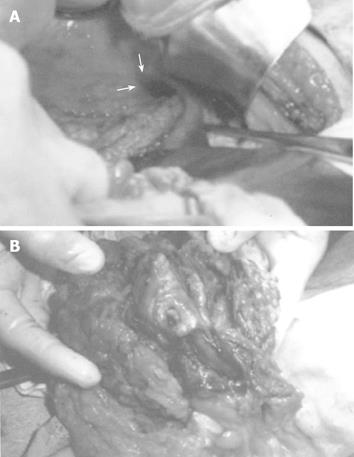
Figure 2 Twisting of greater omentum into one and a half circles and entering into a left inguinal hernia (arrow).
- Citation: Hirano Y, Oyama K, Nozawa H, Hara T, Nakada K, Hada M, Takagi T, Hirano M. Left-sided omental torsion with inguinal hernia. World J Gastroenterol 2006; 12(4): 662-664
- URL: https://www.wjgnet.com/1007-9327/full/v12/i4/662.htm
- DOI: https://dx.doi.org/10.3748/wjg.v12.i4.662


