Copyright
©2006 Baishideng Publishing Group Co.
World J Gastroenterol. Apr 14, 2006; 12(14): 2168-2173
Published online Apr 14, 2006. doi: 10.3748/wjg.v12.i14.2168
Published online Apr 14, 2006. doi: 10.3748/wjg.v12.i14.2168
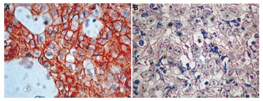
Figure 1 Immunohistochemical staining for positive (A) and negative (B) E-cad expression in diffuse type tumor.
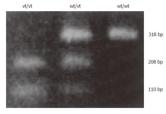
Figure 2 PCR-restriction fragment length polymorphism (RFLP) analysis of genetic polymorphism of the -160 site of the E-cad promoter.
The C/A polymorphism was differentiated by BstEII digestion of PCR products homozygous for the wild-type (high-activity) allele (wt/wt, CC gentoype), heterozygous for the variant (low-activity) allele (wt/vt, CA genotype), and homozygous for the low-activity allele (vt/vt, AA genotype)
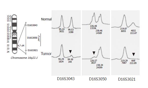
Figure 3 Allelic loss or loss of heterozygosity (LOH) of CDH1/E-cad.
Left panel: E-cad detected by allelic loss or loss of heterozygosity (LOH) of the E-cad locus, reflected by three microsatellite markers (D16S3043, D16SS3050 and D16S3021) at 16q22.1. Right panel: LOH in a representative GC. The locius of markers D16S3043, D16S3050, and D16S3021 were considered to be informative when they were heterozygous in normal tissue (i.e. two alleles were seen), and showed LOH when a 3-fold or greater difference was seen in the relative allele intensity ratio between the tumor and normal DNA (arrow).
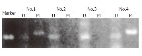
Figure 4 Promoter hypermethylation of the CDH1/E-cad detected by methylation-specific PCR (MSP).
The presence of a visible PCR product in the lanes marked U indicates the presence of an unmethylated allele, while the presence of the product in the lanes marked M indicates the presence of a methylated allele. The intensity of each methylated band was further semi-quantitated, and as shown in the figure, cases 1 and 4 were defined as “hypermethylation”with “+”and “++”, respectively, and cases 2 and 3 were defined as “unmethylation”.
Figure 5 CDH1/E-cad mutation and polymorphism detected by direct DNA sequencing.
Two tumors subjected to DNA sequencing were found to harbor C-to-T transversion in exon 13 (A), resulting in a truncated mutation (Gln to stop condon TAG) and C-to-T transversion in exon 14 (B), resulting in no amino acid change, which was considered to be polymorphism.
- Citation: Liu YC, Shen CY, Wu HS, Hsieh TY, Chan DC, Chen CJ, Yu JC, Yu CP, Harn HJ, Chen PJ, Hsieh CB, Chen TW, Hsu HM. Mechanisms inactivating the gene for E-cadherin in sporadic gastric carcinomas. World J Gastroenterol 2006; 12(14): 2168-2173
- URL: https://www.wjgnet.com/1007-9327/full/v12/i14/2168.htm
- DOI: https://dx.doi.org/10.3748/wjg.v12.i14.2168


