Copyright
©2005 Baishideng Publishing Group Inc.
World J Gastroenterol. Mar 7, 2005; 11(9): 1378-1381
Published online Mar 7, 2005. doi: 10.3748/wjg.v11.i9.1378
Published online Mar 7, 2005. doi: 10.3748/wjg.v11.i9.1378
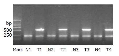
Figure 1 M: marker, DL2000; T1–T4: HCC cancer tissues of four cases, which display a positive band at 585 bp; N1–N4: paracancer tissues; interior reference is seen at 224 bp for all the specimens.
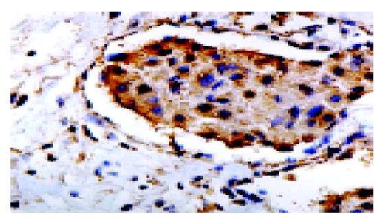
Figure 2 Immunohistochemical staining (brown-yellow) for nm23-H1 in cytoplasm (SP, ×400).
Interstitial cells did not show positive stainings.
- Citation: Liu YB, Gao SL, Chen XP, Peng SY, Fang HQ, Wu YL, Peng CH, Tang Z, Xu B, Wang JW, Deng GL, Li HJ, Feng XD, Qian HR. Expression and significance of heparanase and nm23-H1 in hepatocellular carcinoma. World J Gastroenterol 2005; 11(9): 1378-1381
- URL: https://www.wjgnet.com/1007-9327/full/v11/i9/1378.htm
- DOI: https://dx.doi.org/10.3748/wjg.v11.i9.1378


