Copyright
©2005 Baishideng Publishing Group Inc.
World J Gastroenterol. Mar 7, 2005; 11(9): 1287-1291
Published online Mar 7, 2005. doi: 10.3748/wjg.v11.i9.1287
Published online Mar 7, 2005. doi: 10.3748/wjg.v11.i9.1287
Figure 1 Rectal carcinoma.
A: Target plane of CT perfusion indicates invasive nodule (arrow) and ROI (white round region); B: Corresponding section of specimen shows invasive nodule (five corner star) and examined area (white round region).
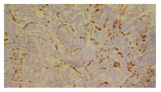
Figure 2 High microvessel density, stained by CD34 (magnification, ×100).
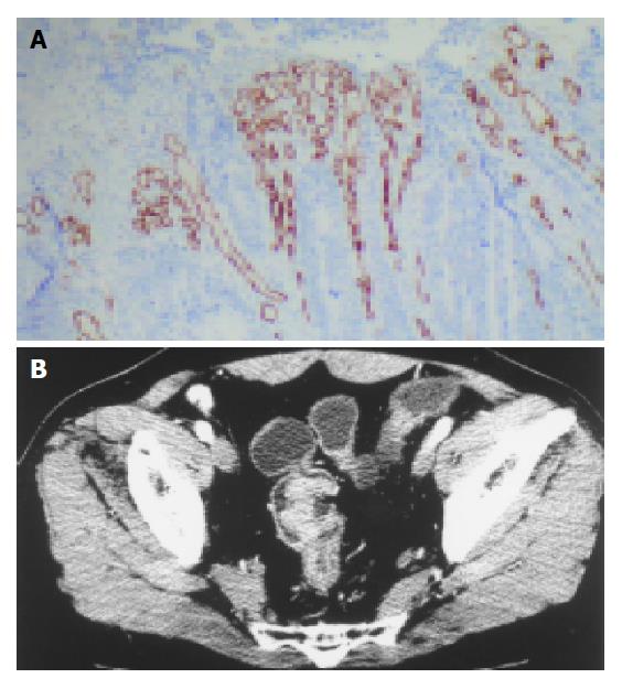
Figure 3 Low microvessel density with high PF.
A: many large vessels with low microvessel density (magnification, ×100); B: Perfusion image shows obvious enhancement of colon lesion with high PF (44.4 mL/min/100 g).
Figure 4 mean±SD for PF for each Dukes’ stage.
- Citation: Li ZP, Meng QF, Sun CH, Xu DS, Fan M, Yang XF, Chen DY. Tumor angiogenesis and dynamic CT in colorectal carcinoma: Radiologic-pathologic correlation. World J Gastroenterol 2005; 11(9): 1287-1291
- URL: https://www.wjgnet.com/1007-9327/full/v11/i9/1287.htm
- DOI: https://dx.doi.org/10.3748/wjg.v11.i9.1287


