Copyright
©2005 Baishideng Publishing Group Inc.
World J Gastroenterol. Feb 28, 2005; 11(8): 1115-1121
Published online Feb 28, 2005. doi: 10.3748/wjg.v11.i8.1115
Published online Feb 28, 2005. doi: 10.3748/wjg.v11.i8.1115
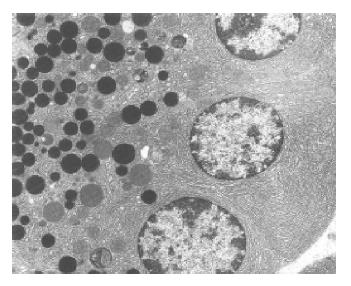
Figure 1 Normal ultrastructural appearance of pancreatic acinar cells.
Control group (C). Original magnification ×3000, bar = 2.5 μm.
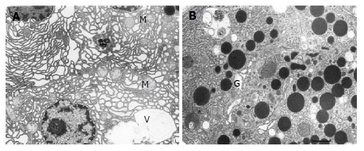
Figure 2 Ultrastructural changes in the pancreatic acinar cells in untreated caerulein-induced AP.
A: Dilated channels of rough endoplasmic reticulum and slightly damaged mitochondria (M) in the cytoplasm of the acinar cells. In the vicinity of nucleus, a vacuole (V) is seen. Original magnification ×3000, bar = 2.5 μm; B: Zymogen granules of different shape and size and dilated cisternae of the Golgi apparatus (G). Original magnification ×7000, bar = 1.1 μm.
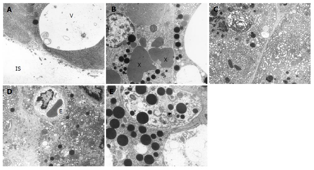
Figure 3 Ultrastructural changes in the pancreatic acinar cells in caerulein-induced AP treated with non-selective (ETA/B) or selective (ETA) ET-1 receptors antagonists.
A: Large vacuole (V) at the base of acinar cell opening into the interstitial space (IS). Group 3 – AP treated with LU 302872 (ETA/B antagonist) at a dose of 10 mg/kg b.w. Original magnification ×4400, bar=1.7 μm; B: Irregular “lakes” of zymogen material (*), large vacuoles and zymogen granules at the base of two acinar cells. Group 3 – AP treated with LU 302872 (ETA/B antagonist) at a dose of 10 mg/kg b.w. Original magnification ×3000, bar=2.5 μm; C: Concentric arrangement, vesicular transformation and disorganization of endoplasmic reticulum. Note some phagosomes (Ph) in the cytoplasm. Group 4 – AP treated with LU 302872 (ETA/B antagonist) at a dose of 20 mg/kg b.w. Original magnification ×3000, bar = 2.5 μm; D: Total disintegration of the acinar cell – its fragments in the interstitial space are seen. The endothelial cells (E) of capillary are swollen. Group 6 – caerulein-induced AP treated with LU 302146 (ETA antagonist) at a dose of 20 mg/kg b.w. Original magnification ×3000, bar = 2.5 μm; E: Autophagous vacuoles containing zymogen granules, fragments of endoplasmic reticulum and other acinar cell organelles. Group 6 – caerulein-induced AP treated with LU 302146 (ETA antagonist ) at a dose of 20 mg/kg b.w. Original magnification ×7000, bar = 1.1 μm.
- Citation: Andrzejewska A, Dlugosz JW, Augustynowicz A. Effect of endothelin-1 receptor antagonists on histological and ultrastructural changes in the pancreas and trypsinogen activation in the early course of caerulein-induced acute pancreatitis in rats. World J Gastroenterol 2005; 11(8): 1115-1121
- URL: https://www.wjgnet.com/1007-9327/full/v11/i8/1115.htm
- DOI: https://dx.doi.org/10.3748/wjg.v11.i8.1115


