Copyright
©The Author(s) 2005.
World J Gastroenterol. Aug 21, 2005; 11(31): 4923-4926
Published online Aug 21, 2005. doi: 10.3748/wjg.v11.i31.4923
Published online Aug 21, 2005. doi: 10.3748/wjg.v11.i31.4923
Figure 1 CT slice of radiotherapy treatment plan in the drainage area (planning system: TMS Helax).
1-7: Coplanar beams. Isodoses: green: 100% = 48.0 Gy; light blue 1: 90% = 43.2 Gy; light blue 2: 80% = 38.4 Gy; light blue 3: 70% = 33.6 Gy; dark blue: 50% = 24.0 Gy. A: PTV (planning target volume): outer red line includes a safety rim. B: Body frame (low density materials to avoid shielding) with positioning marks. C: Vacuum cushion for reproducible fixation of the patient (low density materials to avoid shielding: not visible in CT scan). D: Drainage. E: Radio-opaque fiducials for the coordinate read out in longitudinal direction.
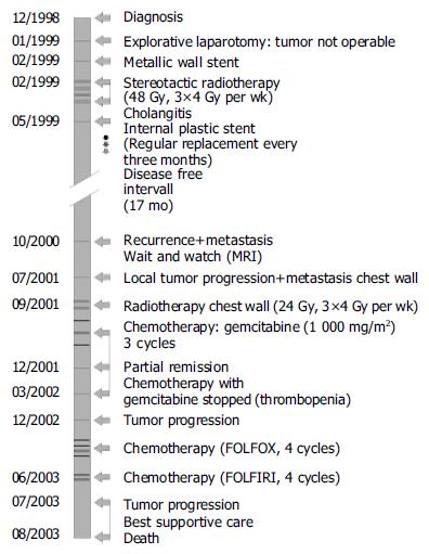
Figure 2 Patient’s treatment course from 12/1998 to 08/2003.
- Citation: Becker G, Momm F, Schwacha H, Hodapp N, Usadel H, Geißler M, Barke A, Schmitt-Gräff A, Henne K, Blum HE. Klatskin tumor treated by inter-disciplinary therapies including stereotactic radiotherapy: A case report. World J Gastroenterol 2005; 11(31): 4923-4926
- URL: https://www.wjgnet.com/1007-9327/full/v11/i31/4923.htm
- DOI: https://dx.doi.org/10.3748/wjg.v11.i31.4923


