Copyright
©2005 Baishideng Publishing Group Inc.
World J Gastroenterol. Apr 21, 2005; 11(15): 2351-2353
Published online Apr 21, 2005. doi: 10.3748/wjg.v11.i15.2351
Published online Apr 21, 2005. doi: 10.3748/wjg.v11.i15.2351
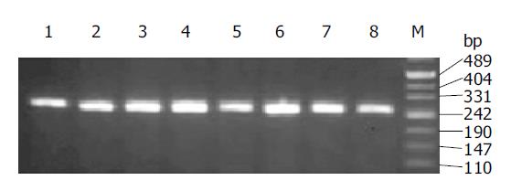
Figure 1 PCR amplification of src exon 12 from genomic DNA of colorectal cancer.
Lanes 1-8: samples; lane M: marker.
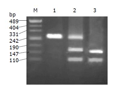
Figure 2 Positive result of No.
73 sample in RFLP analysis. Lane 1: No. 73 para-cancer tissue; lane 2: No. 73 cancer tissue; lane 3: positive control; lane M: DNA marker.
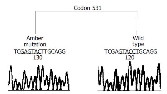
Figure 3 Sequencing of two clones (one wild and the other Amber-mutated) from RFLP-positive PCR products of No.
73 colorectal cancer.
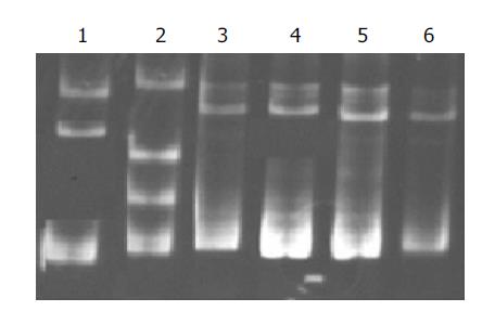
Figure 4 SSCP analysis of src exon 12 fragment amplified from colorectal cancer.
Lane 1: positive control; lane 2: No. 73; lane 3: negative control; lanes 4-6: three negative samples.
- Citation: Tan YX, Wang HT, Zhang P, Yan ZH, Dai GL, Wu MC, Wang HY. c-src activating mutation analysis in Chinese patients with colorectal cancer. World J Gastroenterol 2005; 11(15): 2351-2353
- URL: https://www.wjgnet.com/1007-9327/full/v11/i15/2351.htm
- DOI: https://dx.doi.org/10.3748/wjg.v11.i15.2351


