Copyright
©The Author(s) 2004.
World J Gastroenterol. Feb 1, 2004; 10(3): 455-457
Published online Feb 1, 2004. doi: 10.3748/wjg.v10.i3.455
Published online Feb 1, 2004. doi: 10.3748/wjg.v10.i3.455
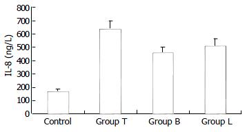
Figure 1 Concentrations of IL-8 in each group (mean ± SD).
Natural interleukin-8 expression was seldom found in HT29 cells of control group. When stimulated by TNF-α (10 ng/ml), HT29 cells secreted a large number of IL-8, and the concentra-tions IL-8 in groups T, B and L were significantly increased than that in control (P < 0.001). However, there was some dif-ference in the concentration of IL-8 among groups T, B and L. There was less interleukin-8 secretion in HT29 cells when preincubated with B. Longum or L. bulgaricus in groups B and L compared with group T (P = 0.002, 0.01, respectively), but there was no difference in concentration of IL-8 between groups B and L (P = 0.068).
Figure 2 Expression of nuclear NF-κB p65 in each group.
Line 1: group T, Line 2: control, Line 3: group L, Line 4: group B.
- Citation: Bai AP, Ouyang Q, Zhang W, Wang CH, Li SF. Probiotics inhibit TNF-α-induced interleukin-8 secretion of HT29 cells. World J Gastroenterol 2004; 10(3): 455-457
- URL: https://www.wjgnet.com/1007-9327/full/v10/i3/455.htm
- DOI: https://dx.doi.org/10.3748/wjg.v10.i3.455


