Copyright
©The Author(s) 2004.
World J Gastroenterol. Nov 1, 2004; 10(21): 3191-3193
Published online Nov 1, 2004. doi: 10.3748/wjg.v10.i21.3191
Published online Nov 1, 2004. doi: 10.3748/wjg.v10.i21.3191
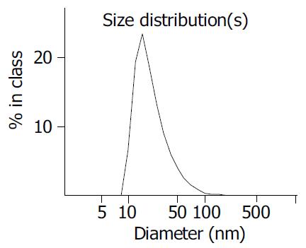
Figure 1 Volume size distribution of TiO2.
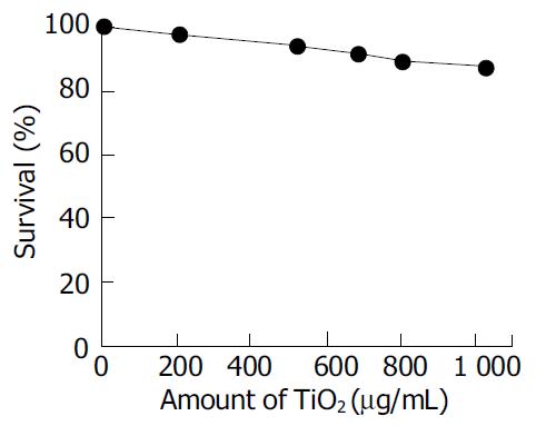
Figure 2 After Ls-174-t cells were incubated in RPMI 1640 medium for 24 h without irradiation, the survival of Ls-174-t cells was shown.
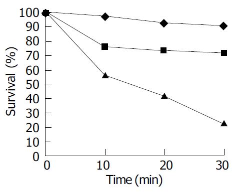
Figure 3 Effect of light and TiO2 on viability of Ls-174-t cells.
(A) TiO2 (1 000 μg/mL) in the dark; (B) no TiO2 in the light; (C) TiO2 (1 000 μg/mL) in the light. Initial cell concentration: 5 × 10⁵ cell/mL, light intensity: 3.7 mW/cm2.
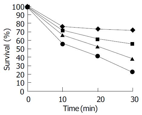
Figure 4 Influence of TiO2 concentration on Ls-174-t cells activity.
(A) 204 μg/mL, (B) 524 μg/mL, (C) 804 μg/mL, (D) 1 029 μg/mL.
-
Citation: Zhang AP, Sun YP. Photocatalytic killing effect of TiO
2 nanoparticles on Ls-174-t human colon carcinoma cells. World J Gastroenterol 2004; 10(21): 3191-3193 - URL: https://www.wjgnet.com/1007-9327/full/v10/i21/3191.htm
- DOI: https://dx.doi.org/10.3748/wjg.v10.i21.3191


