Copyright
©The Author(s) 2004.
World J Gastroenterol. Nov 1, 2004; 10(21): 3182-3184
Published online Nov 1, 2004. doi: 10.3748/wjg.v10.i21.3182
Published online Nov 1, 2004. doi: 10.3748/wjg.v10.i21.3182
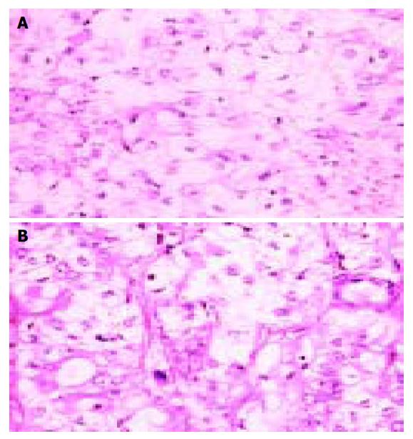
Figure 1 Round tumor cells, with clear cytoplasm, vary greatly in size and shape.
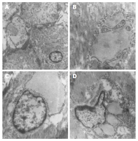
Figure 2 Ultrastructure of gastric leiomyoblastoma.
A: Tu-mor cells with many microfilaments, B: Intracisternal segre-gation could also be found, C: Rough endoplasmic reticulum dilatated as lakes, and protein secretion of intermediate elec-tron density was found in the dilated cisternae, D: Distorted nuclei were found in tumor cells.
- Citation: Huang XF, Wang CM, Pan BR, Dai XW, Fang L, Yang JJ, Yu H, Ren J. Pathological characteristics of gastric leiomyoblastoma. World J Gastroenterol 2004; 10(21): 3182-3184
- URL: https://www.wjgnet.com/1007-9327/full/v10/i21/3182.htm
- DOI: https://dx.doi.org/10.3748/wjg.v10.i21.3182


