Copyright
©The Author(s) 2004.
World J Gastroenterol. Nov 1, 2004; 10(21): 3088-3093
Published online Nov 1, 2004. doi: 10.3748/wjg.v10.i21.3088
Published online Nov 1, 2004. doi: 10.3748/wjg.v10.i21.3088
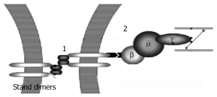
Figure 1 Formation of E-cadherin/catenin complex.
Step1. E-cadherin stand dimers on the two adjacent cells’ surface com-bine together and transfer the signals to catenin complex which is composed of β + α + γ subunits. Step2. The complex promotes actins to contract and enhance the intra-cellular adhesion.
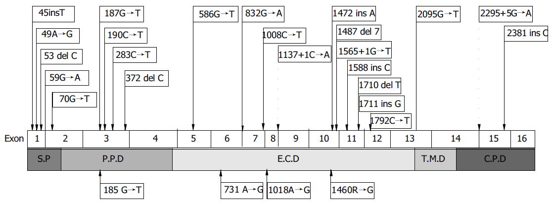
Figure 2 Schematic of CDH1 germline mutations in HGC.
Truncating mutations are shown above the gene and missense mutations below. Arrowhead indicates the status in the dot frame refers to the alteration of intron pointed. S.P: Signal peptide, P.P.D: Protein precursor domain, E.C.D: Extracellular domain, T.M.D: Transmembrane domain and C.P.D: Cytoplastic domain.
- Citation: Wang HD, Ren J, Zhang L. CDH1 germline mutation in hereditary gastric carcinoma. World J Gastroenterol 2004; 10(21): 3088-3093
- URL: https://www.wjgnet.com/1007-9327/full/v10/i21/3088.htm
- DOI: https://dx.doi.org/10.3748/wjg.v10.i21.3088


