Copyright
©The Author(s) 2004.
World J Gastroenterol. Jan 15, 2004; 10(2): 214-217
Published online Jan 15, 2004. doi: 10.3748/wjg.v10.i2.214
Published online Jan 15, 2004. doi: 10.3748/wjg.v10.i2.214
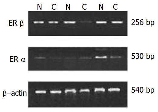
Figure 1 mRNA expression of ERα and ERβ in paired repre-sentative tissues from cancer and adjacent normal mucosa.
C: cancer, N: normal mucosa. RT-PCR result of β-actin was used to show equal loading.
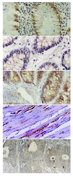
Figure 2 Immunohistochemical staining of ERβ in CRC and nor-mal colonic mucosa.
A: shows the ERβ positive epithelium.×200. B: shows the ERβ positive crypt cell. ×200. C: shows the nuclear and cytoplasmic staining in rectal tubular adenocarcinoma.×200. D: shows the ERβ positive smooth muscle cell and stromal cell. ×400. E shows difuse cytoplasmic staining in CRC. ×400.
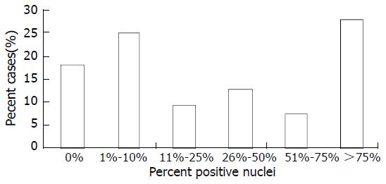
Figure 3 Extent of nuclear ER-β immunoreactivity in 40 cases of CRC.
- Citation: Xie LQ, Yu JP, Luo HS. Expression of estrogen receptor β in human colorectal cancer. World J Gastroenterol 2004; 10(2): 214-217
- URL: https://www.wjgnet.com/1007-9327/full/v10/i2/214.htm
- DOI: https://dx.doi.org/10.3748/wjg.v10.i2.214


