Copyright
©The Author(s) 2004.
World J Gastroenterol. Jan 15, 2004; 10(2): 177-181
Published online Jan 15, 2004. doi: 10.3748/wjg.v10.i2.177
Published online Jan 15, 2004. doi: 10.3748/wjg.v10.i2.177
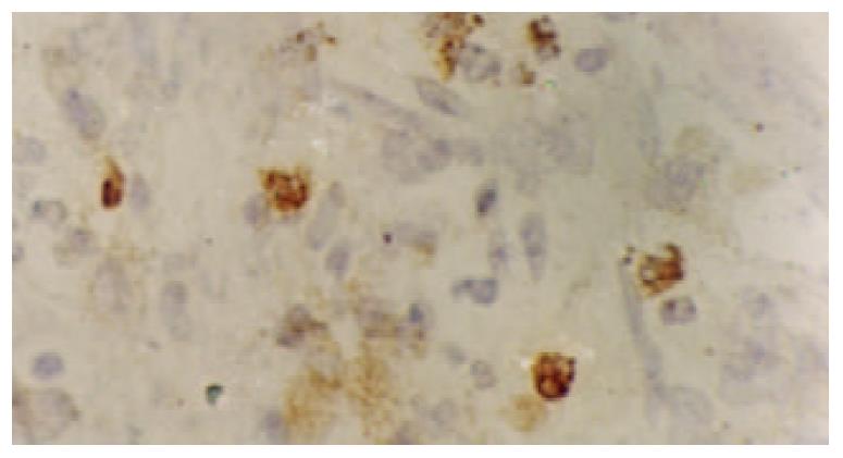
Figure 1 Immunohistochemical detection of NF-κBp65 protein in gastric carcinoma showing cytoplasmic and nuclear staining SP×400.
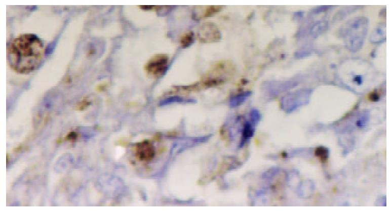
Figure 2 Immunohistochemical detection of hTERT protein in poorly differentiated gastric carcinoma.
A positive reaction was shown in the nuclei of cancer cells. SP×400.
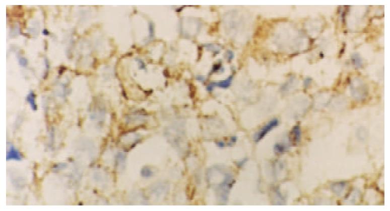
Figure 3 Detection of hTERT mRNA in a mucinous adenocarcinoma.
Most tumor cells displayed strong signals that were localized in the cytoplasm. ISH×400.
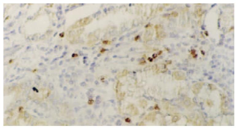
Figure 4 Detection of hTERT mRNA in intestinal metaplasia.
The reaction was localized in the cytoplasm of intestinal meta-plastic cells, activated lymphocytes, as well as replicating cells in basal layer, while the surface epithelia were negative. ISH×100.
- Citation: Wang W, Luo HS, Yu BP. Expression of NF-κB and human telomerase reverse transcriptase in gastric cancer and precancerous lesions. World J Gastroenterol 2004; 10(2): 177-181
- URL: https://www.wjgnet.com/1007-9327/full/v10/i2/177.htm
- DOI: https://dx.doi.org/10.3748/wjg.v10.i2.177


