Copyright
©The Author(s) 2004.
World J Gastroenterol. Oct 1, 2004; 10(19): 2919-2921
Published online Oct 1, 2004. doi: 10.3748/wjg.v10.i19.2919
Published online Oct 1, 2004. doi: 10.3748/wjg.v10.i19.2919
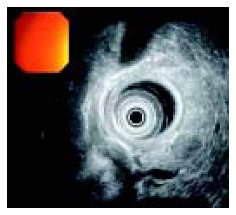
Figure 1 Pancreatic mass (radial endoscopic ultrasound, Olympus device).
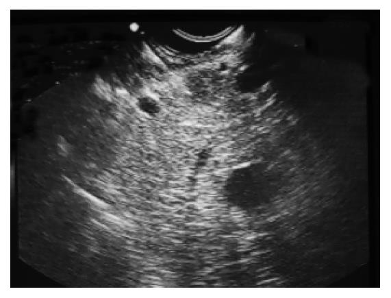
Figure 2 Ultrasound (linear EUS) guided aspiration biopsy of the pancreatic mass.
Figure 3 Groups of epithelial cells with positive cytoplasmic staining on chromogranin (A) and NSE (B) (immunocytochemical analysis).
- Citation: Marisavljevic D, Petrovic N, Milinic N, Cemerikic V, Krstic M, Markovic O, Bilanovic D. An unusual presentation of “silent” disseminated pancreatic neuroendocrine tumor. World J Gastroenterol 2004; 10(19): 2919-2921
- URL: https://www.wjgnet.com/1007-9327/full/v10/i19/2919.htm
- DOI: https://dx.doi.org/10.3748/wjg.v10.i19.2919


