Copyright
©The Author(s) 2004.
World J Gastroenterol. Jul 1, 2004; 10(13): 1885-1889
Published online Jul 1, 2004. doi: 10.3748/wjg.v10.i13.1885
Published online Jul 1, 2004. doi: 10.3748/wjg.v10.i13.1885
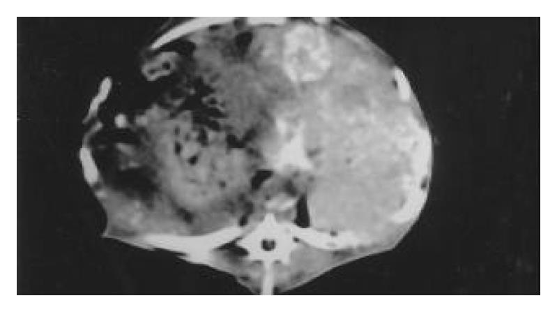
Figure 1 CT scan 2 d following lipiodol infusion.
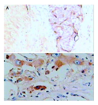
Figure 2 Immunohistochemical staining in hepatic tumor tis-sues obtained from a rabbit underwent TAE.
A: CD31, origi-nal magnification × 200; B: VEGF, original magnification × 400.
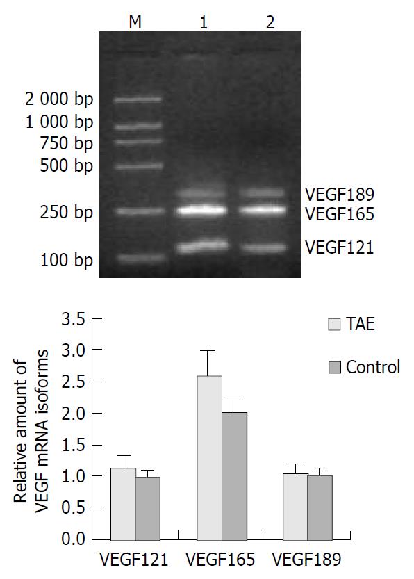
Figure 3 Expression of VEGF mRNA in hepatic tumor tissues.
A: Results of RT-PCR of VEGF mRNA in TAE (lane 1), control (lane 2) and M (molecular mass marker); B: quantification of RT-PCR data.
- Citation: Liao XF, Yi JL, Li XR, Deng W, Yang ZF, Tian G. Angiogenesis in rabbit hepatic tumor after transcatheter arterial embolization. World J Gastroenterol 2004; 10(13): 1885-1889
- URL: https://www.wjgnet.com/1007-9327/full/v10/i13/1885.htm
- DOI: https://dx.doi.org/10.3748/wjg.v10.i13.1885


