Copyright
©The Author(s) 2004.
World J Gastroenterol. Jan 1, 2004; 10(1): 67-72
Published online Jan 1, 2004. doi: 10.3748/wjg.v10.i1.67
Published online Jan 1, 2004. doi: 10.3748/wjg.v10.i1.67
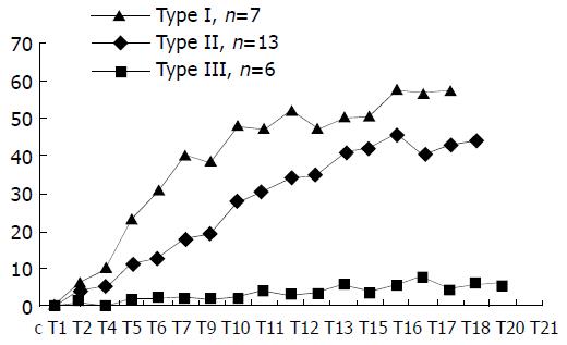
Figure 1 Three patterns (I, II, III) of time-density curve ob-served in HCC patients.
The transverse axis represents the time and the Y-axis represents the peak enhancement value in Hounsfield units.
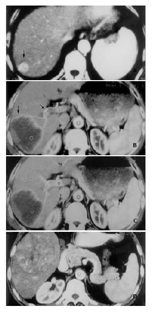
Figure 2 Three types of enhancement morphology depicted in HCC patients.
A: Type A. Marked and homogeneous enhance-ment of the entire HCC lesion in the posterosuperior segment of right hepatic lobe (black arrow). B: Type B. Bright periph-eral ring-like enhancement of HCC lesion on arterial phase image in the right posterosuperior segment (black arrow), and the dilated right hepatic artery (black arrow). C: Portal venous image at the same slice level as in (B). HCC lesion remained hypodense despite obvious enhancement of normal liver pa-renchyma elsewhere. D: Type C. Inhomogeneous patchy en-hancement of HCC lesion in right lower hepatic lobe from ar-terial phase image. Bright dots and linear shadows represent enhanced tumor vessels within the lesion.
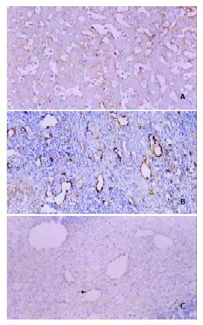
Figure 3 Three patterns of intratumoral MVD distribution revealed by F8RA immunohistochemical staining.
A: Pattern I. Markedly dilated and abundant blood sinusoids with very rich positively stained sinusoidal endothelial cells and scanty tumor interstitium. B: Pattern II. Sinusoids and interstitium abundant and rich in positively stained endothelial cells (black arrow). C: Pattern III. Rich tumor interstitium with few posi-tively stained endothelial cells (white arrow) and scanty blood sinusoids.
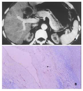
Figure 4 Enhancement of HCC pseudocapsules and pseudocapsular MVD.
A: Marked enhancement of HCC pseudocapsule (hyperdensity) in the medial segment of left hepatic lobe shown by DSCT. Note the similar enhancement pattern of the satellite or daughter lesion in the anterior seg-ment of right hepatic lobe. B: F8RA staining Rich positively stained endothelial cells within tumor pseudocapsule (black arrow) revealed by F8RA staining.
- Citation: Chen WX, Min PQ, Song B, Xiao BL, Liu Y, Ge YH. Single-level dynamic spiral CT of hepatocellular carcinoma: Correlation between imaging features and density of tumor microvessels. World J Gastroenterol 2004; 10(1): 67-72
- URL: https://www.wjgnet.com/1007-9327/full/v10/i1/67.htm
- DOI: https://dx.doi.org/10.3748/wjg.v10.i1.67


