
















Copyright: ©Author(s) 2026.
Artif Intell Gastrointest Endosc. Sep 8, 2026; 7(2): 121644
Published online Sep 8, 2026. doi: 10.37126/aige.121644
Published online Sep 8, 2026. doi: 10.37126/aige.121644

Figure 1 Three snapshots (deidentified operative photograph) of laparoscopic cholecystectomy with dissected hepatocystic triangle and critical view of safety achievement as observed.
A: White light imaging; B: Yellow enhancement (YE); C: Indocyanine green near infrared fluoroscopy modes (ICG NIR). Note improved fat differentiation with YE and precise common bile duct visualization (black arrow) in ICG NIR modes.
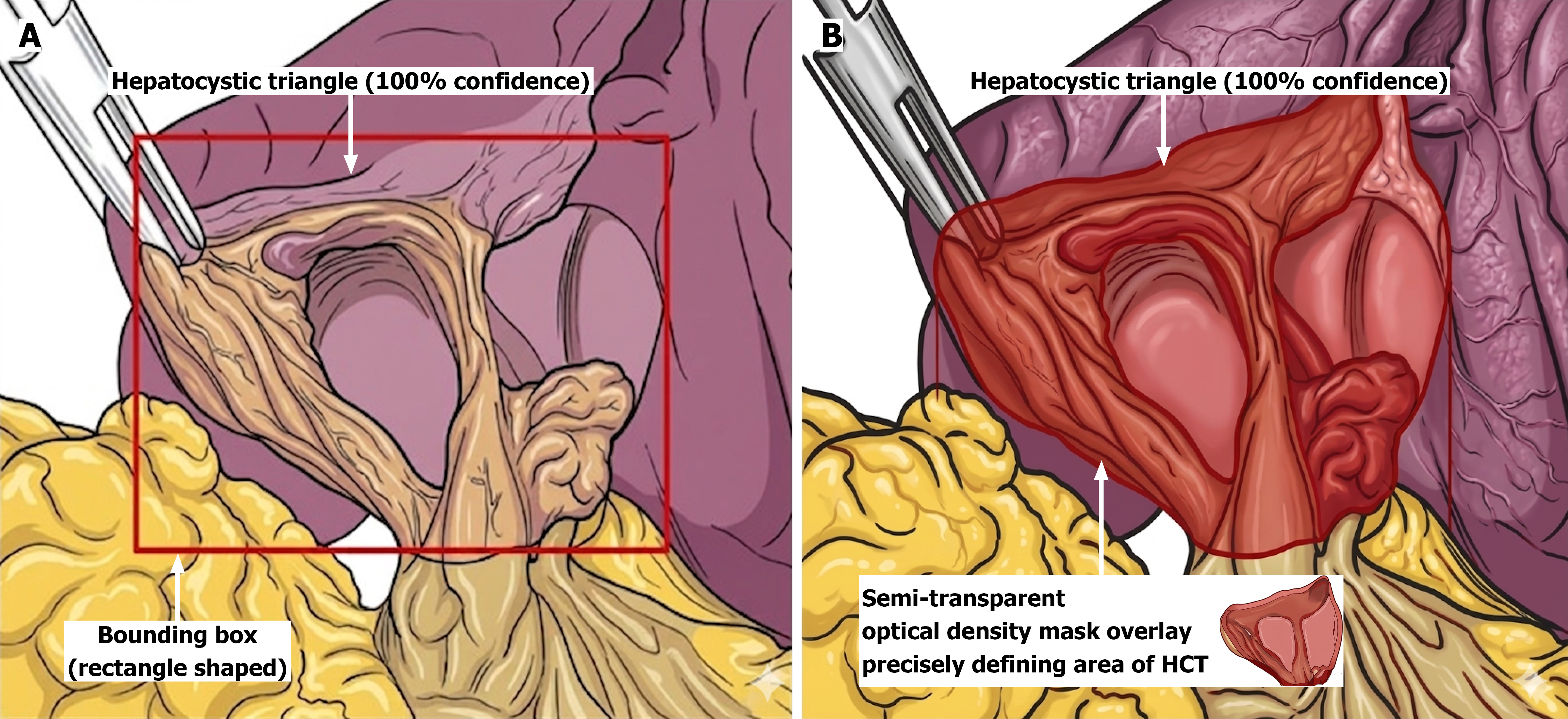
Figure 2 Schematic representation of artificial intelligence-driven anatomic delineation of hepatocystic triangle using bounding box and semi-transparent overlay mask.
A: Initial bounding box method (red rectangle) for broad target area identification and a confidence call out; B: Semantic segmentation further precisely defining the exact region of hepatocystic triangle (HCT) by using a semi-transparent optical density mask overlay.
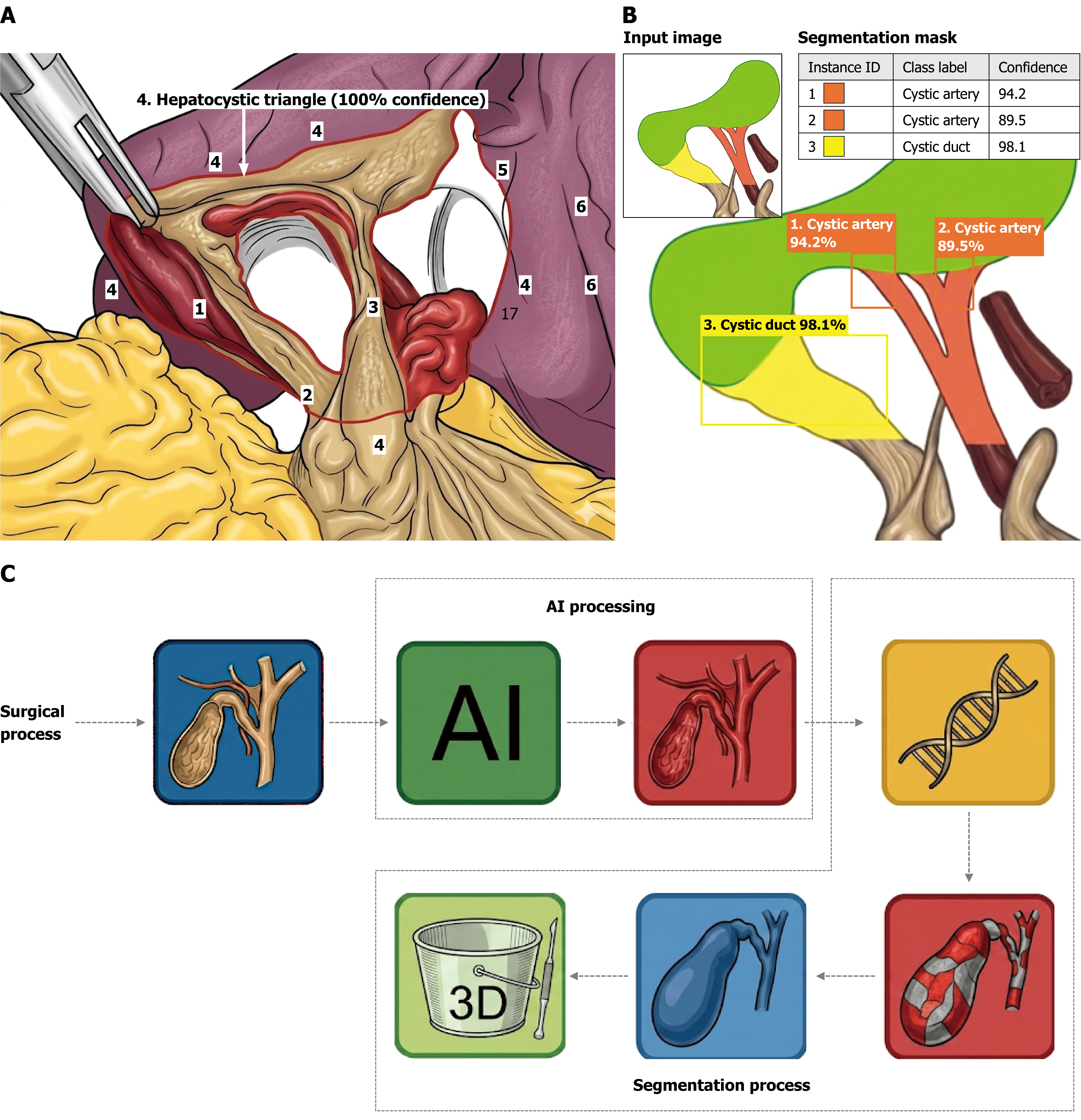
Figure 3 Schematic representation of artificial intelligence processes for anatomical mapping of hepatocystic triangle and critical view of safety assessment using instance segmentation and overview of artificial intelligence workflow pipeline.
A: Integrated artificial intelligence (AI)-enhanced anatomical mapping with transparent mask overlay. This schematic demonstrates the application of landmark-based anatomical mapping used for anatomical segmentation. Numbered identifiers represent key anatomical landmarks that can be used (e.g., 1: Cystic duct-gallbladder junction; 2: Cystic duct-common bile duct junction; 3: Cystic artery; 4: Hepatocystic triangle; 5: Cystic plate; 6: Liver). The figure proposes the concept of using a transparent mask with borders for delineating the hepatocystic triangle (red boundary line). This approach minimizes the obscuration of underlying anatomical textures compared to traditional opaque overlays. The textbox shows the confidence callout of the model in identifying hepatocystic triangle. Numerical labels are illustrative and do not correspond to a specific validated system; B: Instance segmentation for anatomical decomposition and critical view of safety assessment. This schematic illustrates the application of instance segmentation to identify and differentiate individual anatomical structures within the hepatocystic triangle, shown in a spatially decomposed arrangement. Unlike semantic segmentation, which assigns a single class label to all pixels of a given structure type, instance segmentation enumerates discrete anatomical objects individually. This enables automated verification of the critical view of safety, providing an algorithmic baseline to assess the “two-structure rule” (Cystic duct and Cystic artery entering the gallbladder) prior to clip application and ductal division. Numerical labels are illustrative. Clinical application requires surgeon confirmation of model outputs; C: AI pipeline for segmentation and anatomical analysis. Laparoscopic video/still images of the hepatobiliary anatomy are passed to an AI based segmentation model, which extracts hierarchical anatomical features via a convolutional neural network backbone (represented by the double helix motif). The model generates a per-pixel classification map in which each pixel is assigned a discrete tissue or landmark label (represented by the color-coded checkered segmentation mask). Downstream outputs include instance-level identification of individual structures and overlay on the operative image, followed by 3D volumetric reconstruction of the surgical field to support spatial orientation. Icons are schematic representations of computational processes. AI: Artificial intelligence.
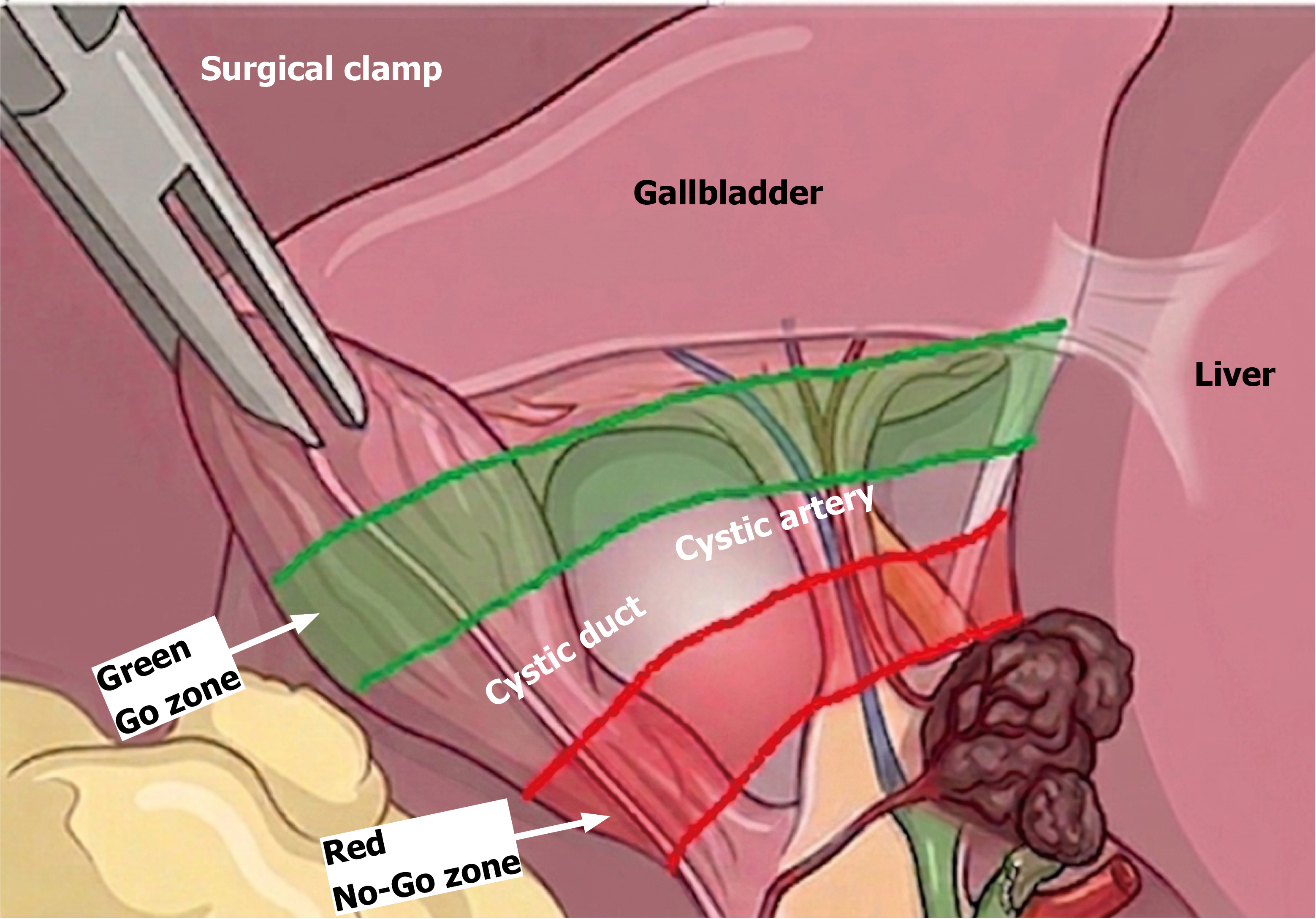
Figure 4
Schematic representation depicting mapping of Safe (Go) and unsafe (No-Go) zones in hepatocystic triangle (please note this is an illustration to show how heat maps are being used to delineate these zones and not an accurately scaled image).
- Citation: Hazrah P, Rautela VS, Sharma V, Mittal S, Madan RR, Sharma D. Improving precision of laparoscopic cholecystectomy: A review on the role of newer intraoperative imaging aids and artificial intelligence. Artif Intell Gastrointest Endosc 2026; 7(2): 121644
- URL: https://www.wjgnet.com/2689-7164/full/v7/i2/121644.htm
- DOI: https://dx.doi.org/10.37126/aige.121644