Copyright
©The Author(s) 2022.
Artif Intell Gastrointest Endosc. Apr 28, 2022; 3(2): 9-15
Published online Apr 28, 2022. doi: 10.37126/aige.v3.i2.9
Published online Apr 28, 2022. doi: 10.37126/aige.v3.i2.9
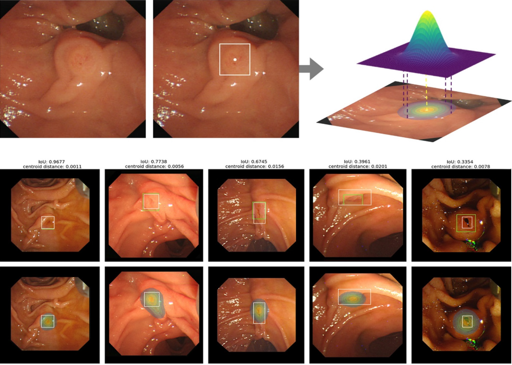
Figure 1 Detection of the location of ampulla of Vater using an artificial intelligence system.
The top image shows the process of obtaining the heat map (in the middle image, the ampulla of Vater is outlined and the centroid identified, and, from that, a pixel-wise soft mask label is created). In the figures below, the artificial intelligence system is being applied to identify the AOV. The white bounding boxes correspond to the ground truth and the green ones and the heat map are created by the AI system. The results reveal an adequate performance of the model even when the IoU is around 30%[12]. Citation: Kim T, Kim J, Choi H, Kim E, Keum B, Jeen Y, Lee H, Chun H, Han S, Kim D, Kwon S, Choo J, Lee J. Artificial intelligence-assisted analysis of endoscopic retrograde cholangio
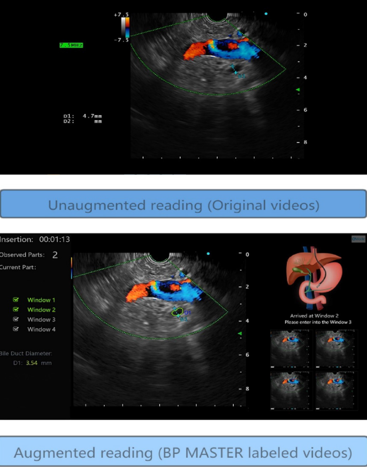
Figure 2 Application of the BP MASTER to endoscopic ultrasound.
In the upper image, there is an image of station 2 (gastric antrum). In the image below, the artificial intelligence system has been applied to identify the station in which the endoscopic ultrasound is located and to identify and delineate the bile duct[29]. Citation: Yao L, Zhang J, Liu J, Zhu L, Ding X, Chen D, Wu H, Lu Z, Zhou W, Zhang L, Xu B, Hu S, Zheng B, Yang Y, Yu H. A deep learning-based system for bile duct annotation and station recognition in linear endoscopic ultrasound. EBioMedicine 2021; 65: 103238. Copyright © The Authors2021. Published by Elsevier B.V.
- Citation: Correia FP, Lourenço LC. Artificial intelligence in the endoscopic approach of biliary tract diseases: A current review. Artif Intell Gastrointest Endosc 2022; 3(2): 9-15
- URL: https://www.wjgnet.com/2689-7164/full/v3/i2/9.htm
- DOI: https://dx.doi.org/10.37126/aige.v3.i2.9


