Copyright
©The Author(s) 2025.
World J Meta-Anal. Jun 18, 2025; 13(2): 100483
Published online Jun 18, 2025. doi: 10.13105/wjma.v13.i2.100483
Published online Jun 18, 2025. doi: 10.13105/wjma.v13.i2.100483
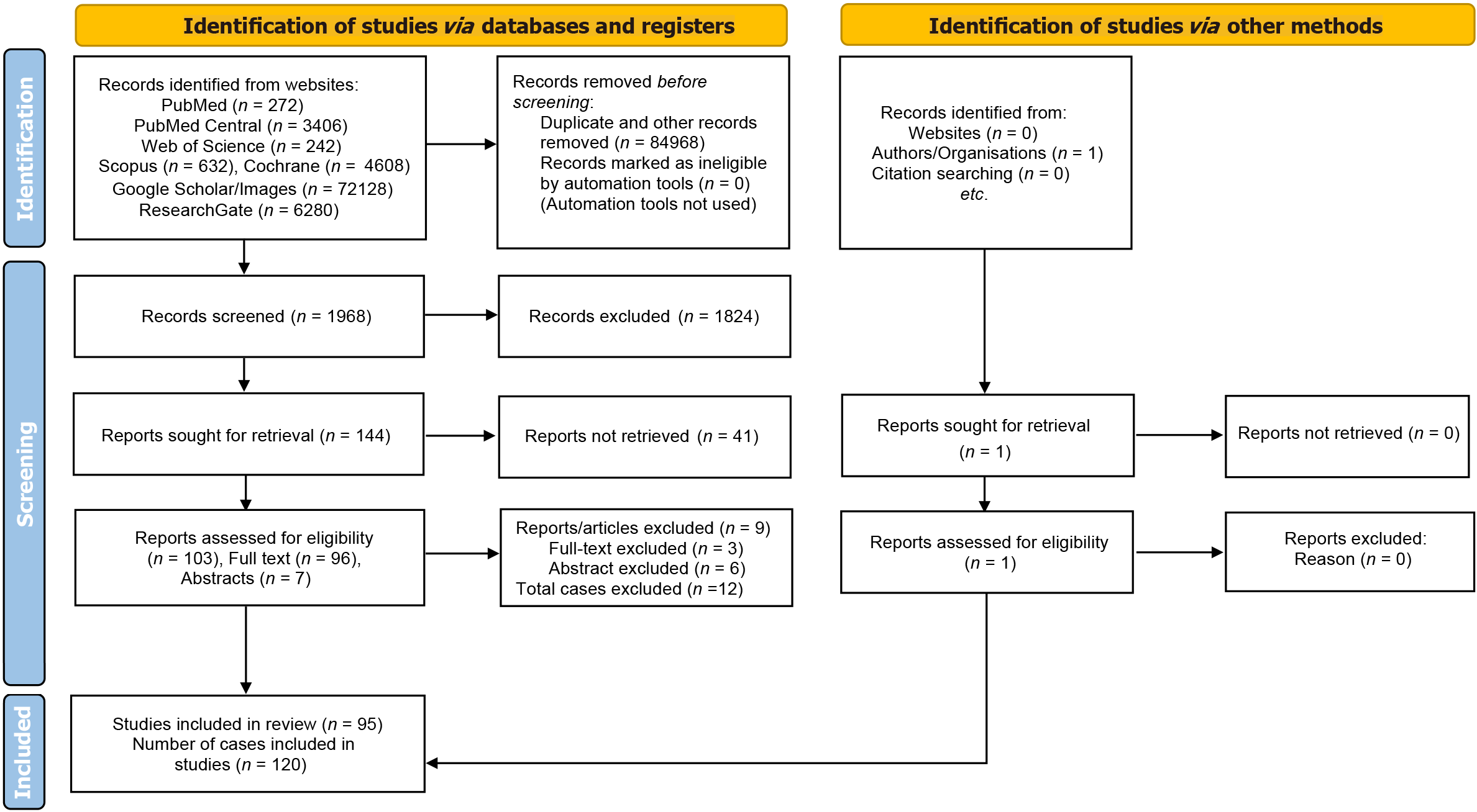
Figure 1
Preferred Reporting Items for Systematic Reviews and Meta Analyses flow diagram for literature search for the systematic review of migration of the distal ventriculoperitoneal shunt catheter into the scrotum.
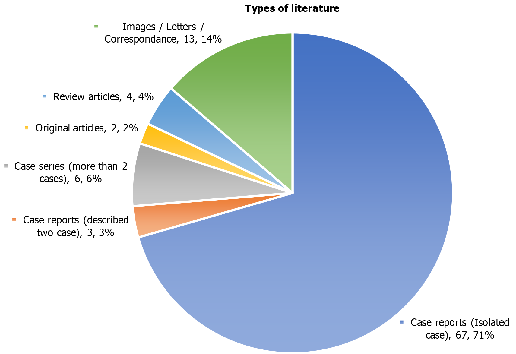
Figure 2
Types of literature (manuscripts) included in the systematic review (n = 95).
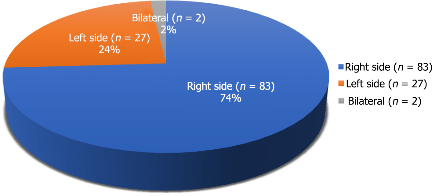
Figure 3
In children, the side of migration of the distal ventriculoperitoneal shunt catheter into the scrotum (n = 112).
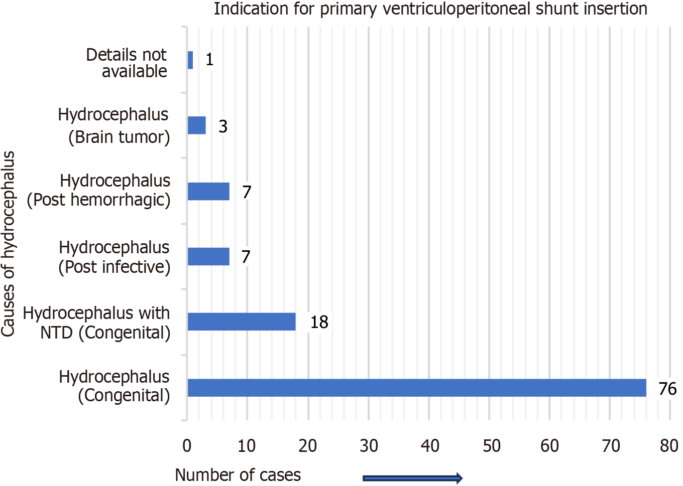
Figure 4 In children, the indication (causes of hydrocephalus) for primary ventriculoperitoneal shunt insertion (n = 112).
NTD: Neural tube defect.
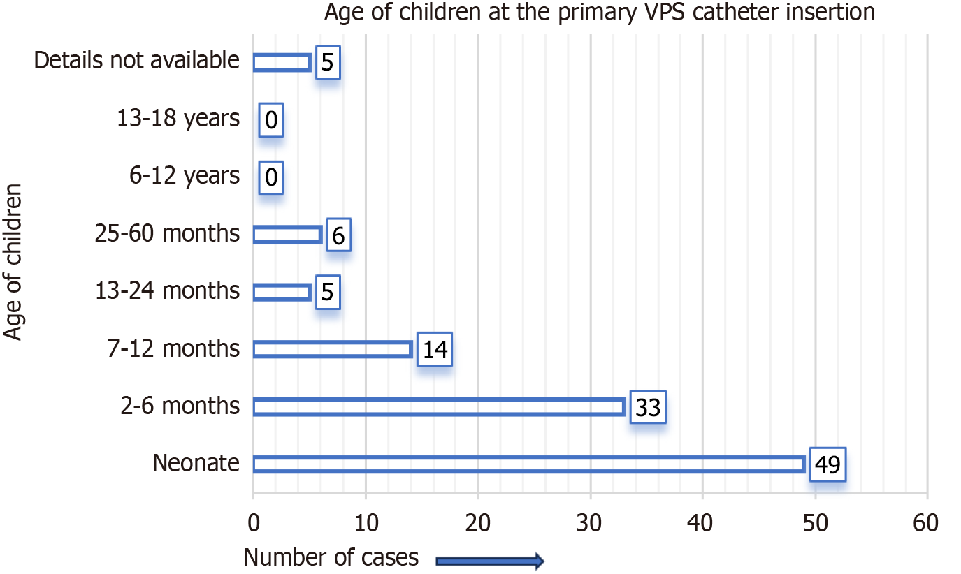
Figure 5 In children, their age at the time of primary ventriculoperitoneal shunt catheter insertion (n = 112).
VPS: Ventriculoperitoneal shunt.
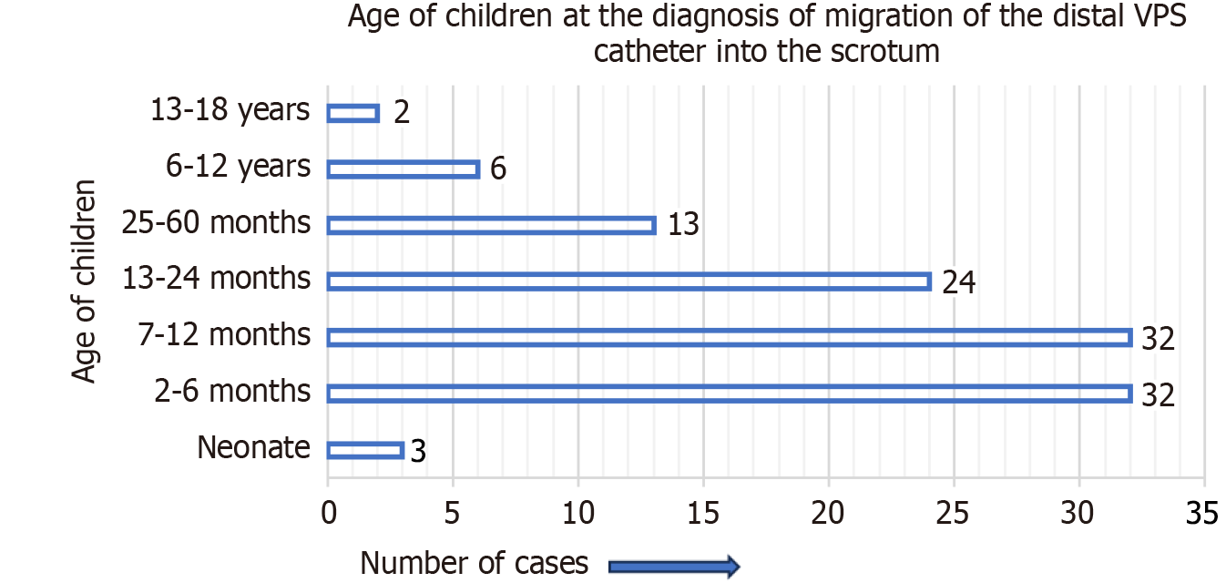
Figure 6 In children, their age at the time of diagnosis of migration of the distal ventriculoperitoneal shunt catheter into the scrotum (n = 112).
VPS: Ventriculoperitoneal shunt.
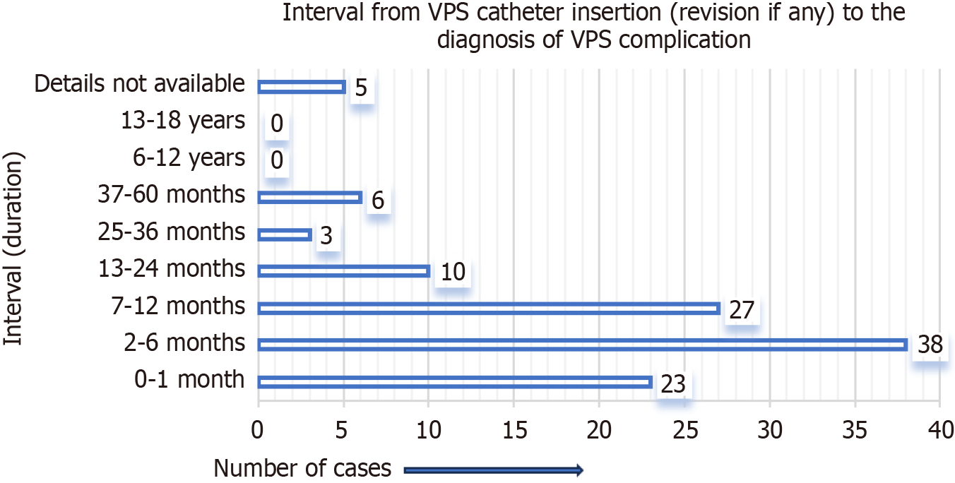
Figure 7 In children, the interval from primary ventriculoperitoneal shunt catheter insertion (revision, if any) to the diagnosis of migration of the distal ventriculoperitoneal shunt catheter into the scrotum (n = 112).
VPS: Ventriculoperitoneal shunt.
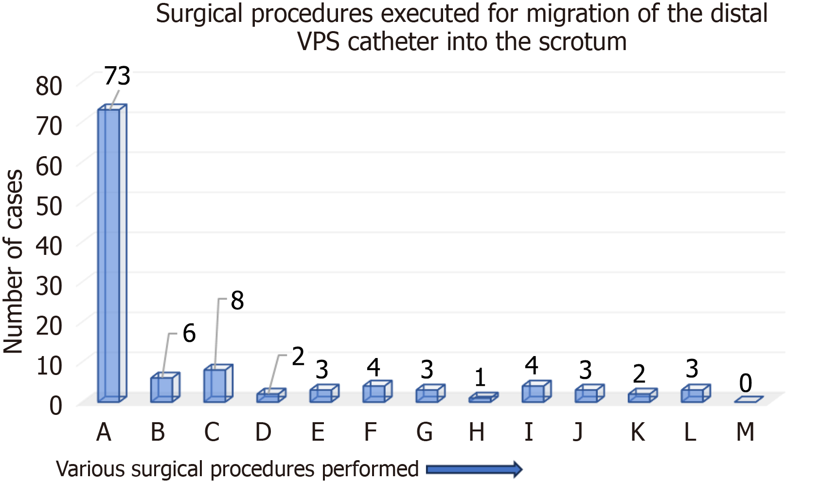
Figure 8 In children, surgical procedures executed for migration of the distal ventriculoperitoneal shunt catheter into the scrotum (n = 112).
A: Reposition of migrated distal ventriculoperitoneal shunt catheter into the peritoneal cavity and inguinal herniotomy; B: Reposition (spontaneous) of migrated distal ventriculoperitoneal shunt catheter into the peritoneal cavity and inguinal herniotomy; C: Removal of part of migrated distal ventriculoperitoneal shunt catheter, repositioning of the remaining peritoneal catheter into the peritoneal cavity and inguinal herniotomy; D: Removal of part of migrated distal ventriculoperitoneal shunt catheter, repositioning of the remaining peritoneal catheter into the peritoneal cavity without inguinal herniotomy; E: Removal of migrated/abandoned distal ventriculoperitoneal shunt catheter with repositioning of working distal ventriculoperitoneal shunt catheter into the peritoneal cavity and inguinal herniotomy; F: Removal of migrated/abandoned distal ventriculoperitoneal shunt catheter and inguinal herniotomy; G: Removal of the entire ventriculoperitoneal shunt catheter alone; H: Removal of the entire ventriculoperitoneal shunt catheter and inguinal herniotomy; I: Removal of entire ventriculoperitoneal shunt catheter with re-ventriculoperitoneal shunt insertion/ventriculoperitoneal shunt revision and inguinal herniotomy; J: Surgical therapy not required for the migrated distal ventriculoperitoneal shunt catheter; K: Surgical therapy not done for the migrated distal ventriculoperitoneal shunt catheter; L: Surgical therapy details not available for the migrated distal ventriculoperitoneal shunt catheter; M: Other surgical procedures for the migrated distal ventriculoperitoneal shunt catheter; VPS: Ventriculoperitoneal shunt.
- Citation: Ghritlaharey RK. Why the migration of peritoneal shunt catheter into the scrotum occurs more in children: A systematic literature review. World J Meta-Anal 2025; 13(2): 100483
- URL: https://www.wjgnet.com/2308-3840/full/v13/i2/100483.htm
- DOI: https://dx.doi.org/10.13105/wjma.v13.i2.100483


