Copyright
©The Author(s) 2017.
World J Methodol. Dec 26, 2017; 7(4): 139-147
Published online Dec 26, 2017. doi: 10.5662/wjm.v7.i4.139
Published online Dec 26, 2017. doi: 10.5662/wjm.v7.i4.139
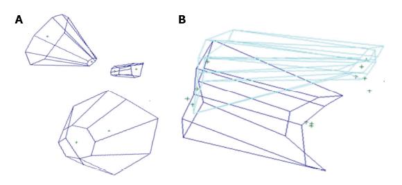
Figure 4 Volume rendering with Maya®.
A: Visual rendering of three different funnels with the quantification method based on the commercially-available digitizer or navigation system and 3-D rendering software (Maya®); B: 3D rendering with the Storz navigation system for coordinate collection and Maya® software for 3D rendering of endonasal (light blue) and transoral (dark blue) approaches to the anterior craniovertebral junction.
- Citation: Doglietto F, Qiu J, Ravichandiran M, Radovanovic I, Belotti F, Agur A, Zadeh G, Fontanella MM, Kucharczyk W, Gentili F. Quantitative comparison of cranial approaches in the anatomy laboratory: A neuronavigation based research method. World J Methodol 2017; 7(4): 139-147
- URL: https://www.wjgnet.com/2222-0682/full/v7/i4/139.htm
- DOI: https://dx.doi.org/10.5662/wjm.v7.i4.139


