Copyright
©The Author(s) 2016.
World J Methodol. Mar 26, 2016; 6(1): 126-132
Published online Mar 26, 2016. doi: 10.5662/wjm.v6.i1.126
Published online Mar 26, 2016. doi: 10.5662/wjm.v6.i1.126
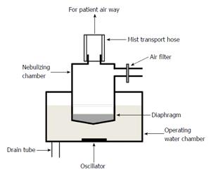
Figure 1 Components and structure of an ultrasonic nebulizer.
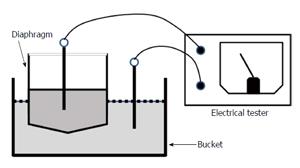
Figure 2 Scheme for discovering diaphragm damage using an electrical tester.
A low concentration of detergent is added to the water in the diaphragm and bucket. Diaphragm breakage or pinholes are detected by measuring the electricity between the diaphragm and bucket.
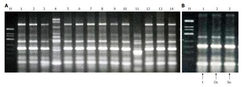
Figure 3 DNA fingerprints of strains determined by random amplified polymorphic DNA assay.
A: Isolates from each patient (1-14); B: Isolates from nebulizer components. t: Nebulizer drain tube; Os: Oscillator; So: Nebulizer solution; M: DNA size marker.
- Citation: Ida Y, Ohnishi H, Araki K, Saito R, Kawai S, Watanabe T. Efficient management and maintenance of ultrasonic nebulizers to prevent microbial contamination. World J Methodol 2016; 6(1): 126-132
- URL: https://www.wjgnet.com/2222-0682/full/v6/i1/126.htm
- DOI: https://dx.doi.org/10.5662/wjm.v6.i1.126


