Copyright
©The Author(s) 2015.
World J Methodol. Sep 26, 2015; 5(3): 144-148
Published online Sep 26, 2015. doi: 10.5662/wjm.v5.i3.144
Published online Sep 26, 2015. doi: 10.5662/wjm.v5.i3.144
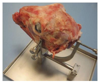
Figure 1 The head holder.
Six sideward-screws serve to fix the lamb's head in desired position while dissecting.
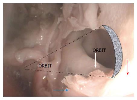
Figure 2 Endoscopic view to the left maxillary complex.
The perpendicular crest (white arrow) divides the cavity within the maxilla into maxillary sinus proper (red arrow) and palatine sinus (blue arrow).
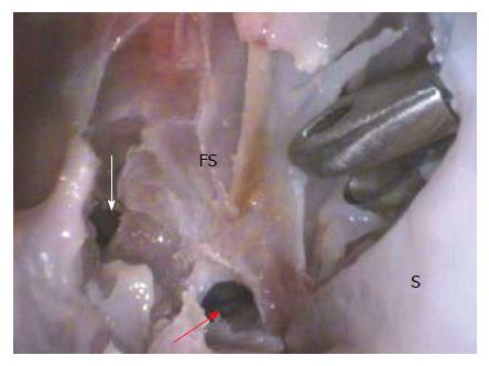
Figure 3 Endoscopic 30° view at the region of the bottom of the frontal sinus.
The tip of the Kerisson’s punch juts from the left nasal cavity through the artificially made septal (S) defect. Red and white arrows indicate the frontal sinus lateral and anterior cells. FS: Frontal sinus.
- Citation: Skitarelić N, Mladina R. Lamb’s head: The model for novice education in endoscopic sinus surgery. World J Methodol 2015; 5(3): 144-148
- URL: https://www.wjgnet.com/2222-0682/full/v5/i3/144.htm
- DOI: https://dx.doi.org/10.5662/wjm.v5.i3.144


