Copyright
©The Author(s) 2019.
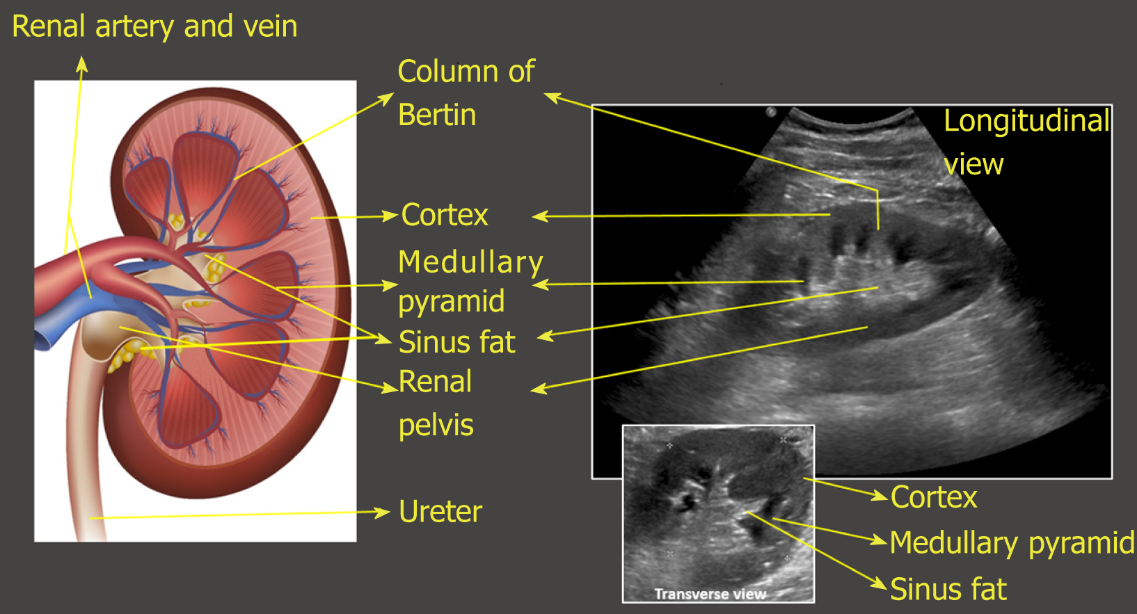
Figure 8 Illustration of longitudinal section of the kidney with corresponding ultrasound image.
Transverse view of the kidney is also shown. Note that the most echogenic part of the kidney is sinus fat, and calyces and ureter are not usually visible unless distended. Renal pelvis is hypoechoic but not usually “black” (anechoic) unless there is hydronephrosis.
- Citation: Koratala A, Bhattacharya D, Kazory A. Point of care renal ultrasonography for the busy nephrologist: A pictorial review. World J Nephrol 2019; 8(3): 44-58
- URL: https://www.wjgnet.com/2220-6124/full/v8/i3/44.htm
- DOI: https://dx.doi.org/10.5527/wjn.v8.i3.44


