Copyright
©The Author(s) 2015.
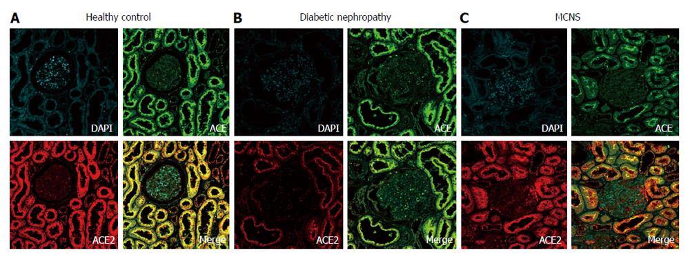
Figure 2 Images obtained with confocal microscopy of triple immunofluorescence staining of angiotensin-converting enzyme (green), angiotensin-converting enzyme 2 (red), and nuclei (DAPI; blue) in kidney specimens.
In healthy subjects, marked co-localization of ACE and ACE2 (yellow) was observed in the apical brush borders of the proximal tubules (A). Both ACE and ACE2 were also present in the glomeruli, but the glomeruli exhibited weaker staining than the proximal tubules (A). In diabetic kidneys, stronger ACE expression and weaker ACE2 expression were detected in the proximal tubules, and no marked colocalization of ACE and ACE2 was observed (B); however, similar ACE and ACE2 staining patterns were seen in patients with minimal change nephrotic syndrome (MCNS) and healthy controls (C). Adapted with permission from Mizuiri et al[33]. ACE: Angiotensin-converting enzyme.
- Citation: Mizuiri S, Ohashi Y. ACE and ACE2 in kidney disease. World J Nephrol 2015; 4(1): 74-82
- URL: https://www.wjgnet.com/2220-6124/full/v4/i1/74.htm
- DOI: https://dx.doi.org/10.5527/wjn.v4.i1.74


