Copyright
©The Author(s) 2018.
World J Nephrol. Dec 17, 2018; 7(8): 155-160
Published online Dec 17, 2018. doi: 10.5527/wjn.v7.i8.155
Published online Dec 17, 2018. doi: 10.5527/wjn.v7.i8.155
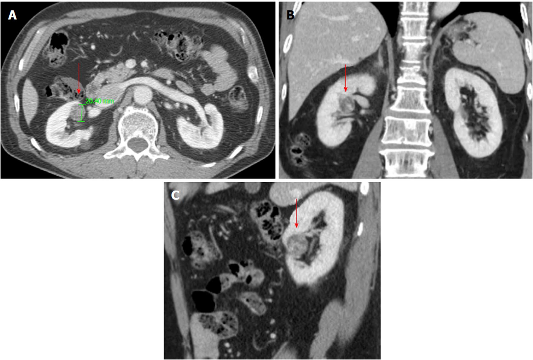
Figure 1 Computed tomography findings of clear cell papillary renal cell carcinoma.
A 2-cm mass with an area of irregular enhancement is visible in the right kidney. A: Axial view; B: Coronal view; C: Sagittal view.
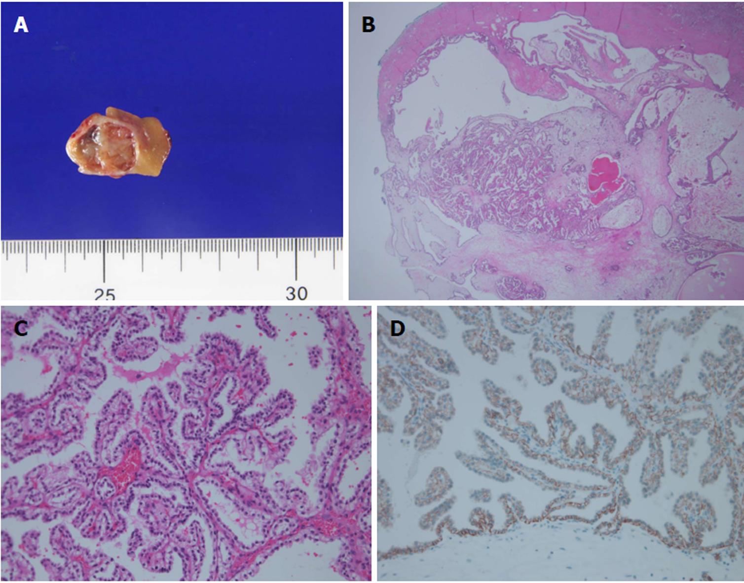
Figure 2 Photomicrographs of clear cell papillary renal cell carcinoma.
A: Gross findings; B: H and E: The tumor showed papillary, cystic and tubular patterns in low powered magnification (original magnification × 12.5); C: The tumor is composed of clear cells with uniform nuclei typically showing a linear arrangement away from the basal membrane (original magnification × 200); D: The typical immunohistochemical staining of carbonic anhydrase IX. The staining pattern is a cup-like distribution typical feature of clear cell papillary renal cell carcinoma (original magnification × 200).
- Citation: Kim SH, Kwon WA, Joung JY, Seo HK, Lee KH, Chung J. Clear cell papillary renal cell carcinoma: A case report and review of the literature. World J Nephrol 2018; 7(8): 155-160
- URL: https://www.wjgnet.com/2220-6124/full/v7/i8/155.htm
- DOI: https://dx.doi.org/10.5527/wjn.v7.i8.155


