Copyright
©The Author(s) 2018.
World J Nephrol. Nov 24, 2018; 7(7): 143-147
Published online Nov 24, 2018. doi: 10.5527/wjn.v7.i7.143
Published online Nov 24, 2018. doi: 10.5527/wjn.v7.i7.143
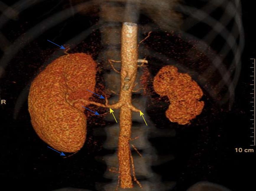
Figure 1 Three-dimensional computed tomography reconstruction.
Extensive collateral blood supply to the right kidney (blue arrows) and origins of the renal arterial stenosis (yellow arrows) are shown.
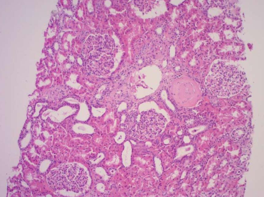
Figure 2 Results of the kidney biopsy.
Normal appearing tissue (haematoxylin and eosin stain, at 100 × high power field magnification).
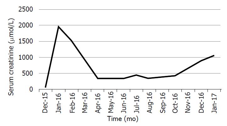
Figure 3 The patient’s serum creatinine levels over time.
- Citation: Chothia MY, Davids MR, Bhikoo R. Awakening the sleeping kidney in a dialysis-dependent patient with fibromuscular dysplasia: A case report and review of literature. World J Nephrol 2018; 7(7): 143-147
- URL: https://www.wjgnet.com/2220-6124/full/v7/i7/143.htm
- DOI: https://dx.doi.org/10.5527/wjn.v7.i7.143


