Copyright
©2014 Baishideng Publishing Group Inc.
World J Nephrol. Aug 6, 2014; 3(3): 118-121
Published online Aug 6, 2014. doi: 10.5527/wjn.v3.i3.118
Published online Aug 6, 2014. doi: 10.5527/wjn.v3.i3.118
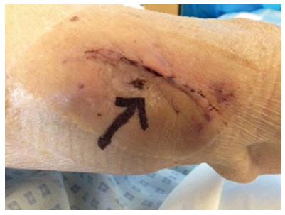
Figure 1 A 3 mm size scab over the arteriovenous fistula (black arrow).
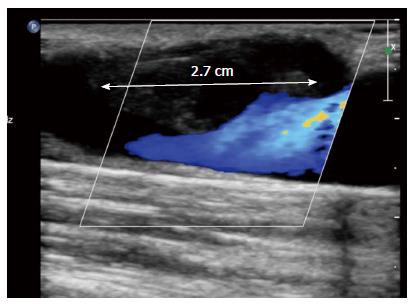
Figure 2 Colour Doppler scan showing a 2.
7 cm long thrombus (white arrow) partially occluding the lumen of the arteriovenous fistula.
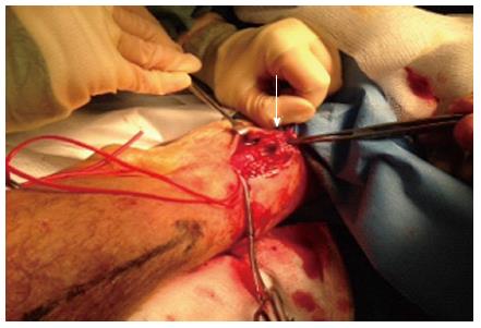
Figure 3 A 2.
5 cm long defect on the anterior wall of the vein (white arrow).
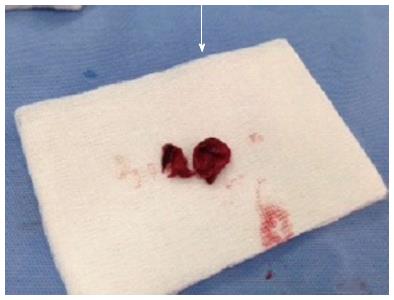
Figure 4 Thrombus removed from the defect in the vessel wall.
- Citation: Shrestha B, Boyes S, Brown P. Innocuous-looking skin scab over an arteriovenous fistula: Case report and literature review. World J Nephrol 2014; 3(3): 118-121
- URL: https://www.wjgnet.com/2220-6124/full/v3/i3/118.htm
- DOI: https://dx.doi.org/10.5527/wjn.v3.i3.118


