Copyright
©2014 Baishideng Publishing Group Inc.
World J Nephrol. Aug 6, 2014; 3(3): 114-117
Published online Aug 6, 2014. doi: 10.5527/wjn.v3.i3.114
Published online Aug 6, 2014. doi: 10.5527/wjn.v3.i3.114
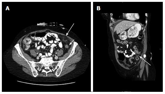
Figure 1 Computed tomography.
A: Axial computed tomography (CT) scan of the abdomen showing a fat density area with some surrounding inflammation (large arrow) anterior to the descending colon (small arrow) in the left iliac fossa just below the abdominal wall at the site of epiploic appendagitis; B: A coronal oblique CT image showing the fat density area with surrounding inflammation (arrow) adjacent to the descending colon.
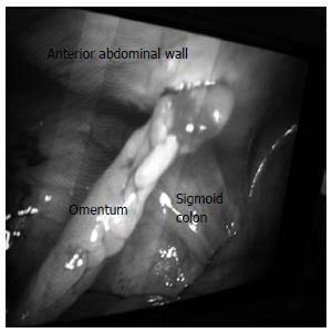
Figure 2 Laparoscopic appearance showing the omentum and anterior surface of the sigmoid colon adherent to the peritoneum lining the anterior abdominal wall in the left iliac fossa.
- Citation: Shrestha B, Hampton J. Recurrent epiploic appendagitis and peritoneal dialysis: A case report and literature review. World J Nephrol 2014; 3(3): 114-117
- URL: https://www.wjgnet.com/2220-6124/full/v3/i3/114.htm
- DOI: https://dx.doi.org/10.5527/wjn.v3.i3.114


