Copyright
©The Author(s) 2023.
World J Virol. Dec 25, 2023; 12(5): 296-308
Published online Dec 25, 2023. doi: 10.5501/wjv.v12.i5.296
Published online Dec 25, 2023. doi: 10.5501/wjv.v12.i5.296
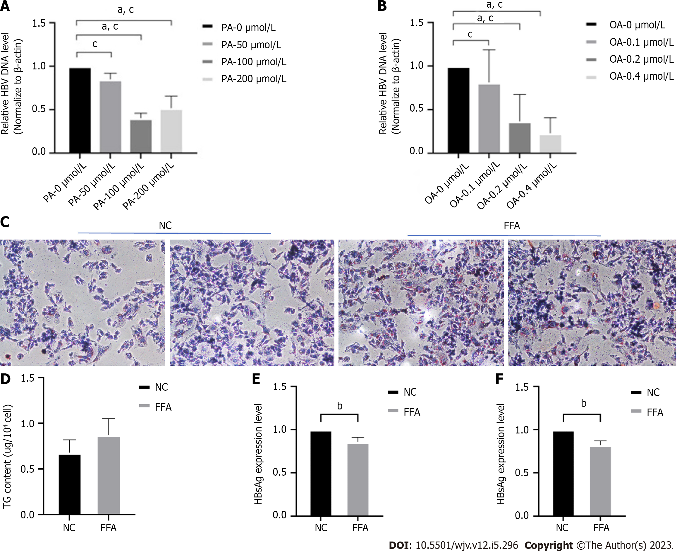
Figure 1 In vitro, high lipid cases promote lipid droplet formation and inhibit chronic hepatitis B virus deoxyribonucleic acid replication and the choreographing of related antibodies.
A and B: HepG2.2.15 cells were stimulated with different concentrations of palmitic acid (PA) and oleic acid (OA) for 48 h, and the expression of chronic hepatitis B virus deoxyribonucleic acid was detected; C: 0.2 mmol/L concentration of OA and 100 μmol/L concentration of PA were applied to stimulate HepG2.2.15 cells for 48 h, and the intracellular lipid droplet formation was detected by applying oil red O staining method; D: Detection of intracellular triglyceride content in both groups; E and F: After applying free fatty acids stimulation for 48 h, the levels of HBsAg and HBeAg secreted by the two groups of cells were detected by the ELISA method, respectively. aP < 0.05, bP < 0.01, cP < 0.001. NC: Nucleocapsid protein; FFA: Free fatty acids.
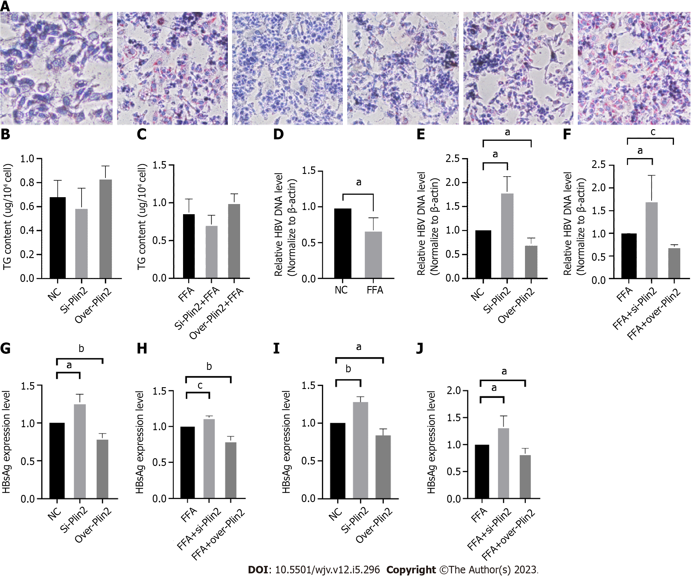
Figure 2 To investigate the role of Plin2 in the effect of high-lipid conditions on chronic hepatitis B virus.
A: Oil red O staining to observe lipid droplet formation; B and C: Triglyceride (TG) assay kit was applied to detect the TG content of each group; D-F: Quantitative polymerase chain reaction method was applied to detect the chronic hepatitis B virus deoxyribonucleic acid content in each group; G-J: HBsAg and HBeAg levels were detected in each group by the ELISA method. aP < 0.05, bP < 0.01, cP < 0.001.
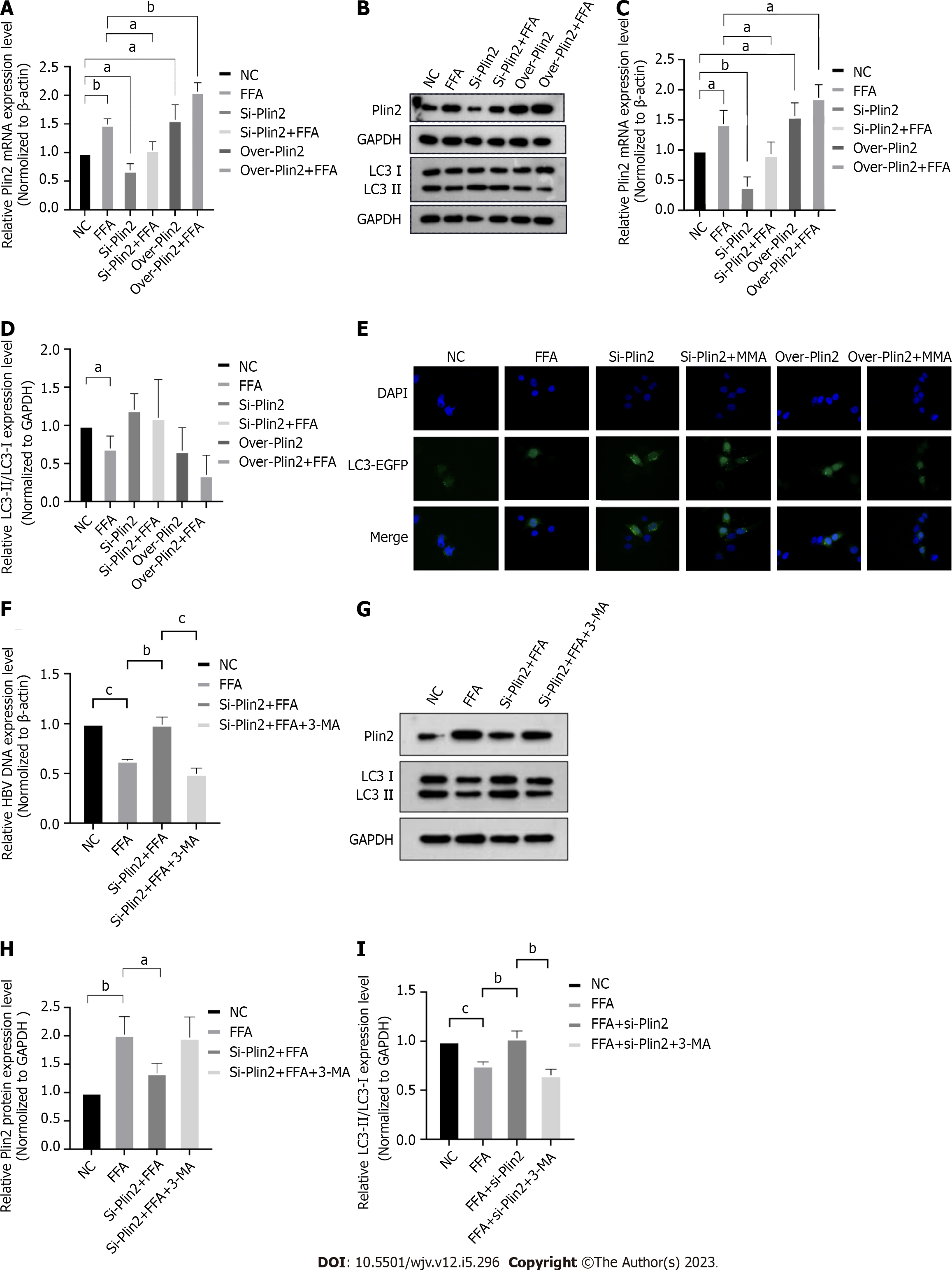
Figure 3 Abnormal lipid metabolism affects chronic hepatitis B virus deoxyribonucleic acid replication through the Plin2-autophagy-related pathway.
A: Quantitative polymerase chain reaction (q-PCR) method was applied to detect Plin2 mRNA levels; B-D: Western blot to detect the expression of Plin2 and LC3 in each group and detect the grayscale value; E: GFP-LC3 formation was observed under the fluorescence microscope; F: Chronic hepatitis B virus deoxyribonucleic acid levels were detected by applying q-PCR; G-I: Western blot was performed to detect the expression of Plin2 and LC3 in each group and to detect the grayscale values. aP < 0.05, bP < 0.01, cP < 0.001.
- Citation: Wang C, Gao XY, Han M, Jiang MC, Shi XY, Pu CW, Du X. Perilipin2 inhibits the replication of hepatitis B virus deoxyribonucleic acid by regulating autophagy under high-fat conditions. World J Virol 2023; 12(5): 296-308
- URL: https://www.wjgnet.com/2220-3249/full/v12/i5/296.htm
- DOI: https://dx.doi.org/10.5501/wjv.v12.i5.296


