Copyright
©The Author(s) 2018.
World J Transplant. Apr 24, 2018; 8(2): 38-43
Published online Apr 24, 2018. doi: 10.5500/wjt.v8.i2.38
Published online Apr 24, 2018. doi: 10.5500/wjt.v8.i2.38
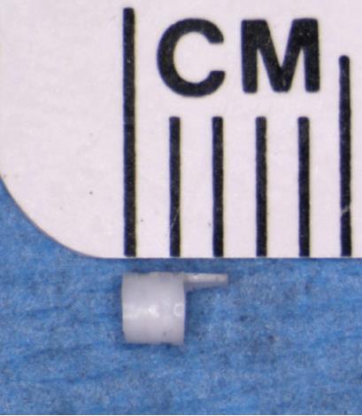
Figure 1 The anastomotic cuff.
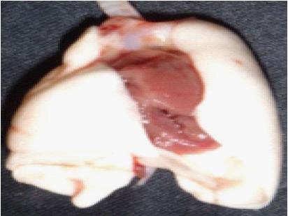
Figure 2 Donor pneumonectomy specimen.
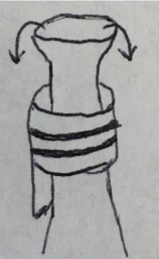
Figure 3 Eversion of the donor vein over the anastomotic cuff.
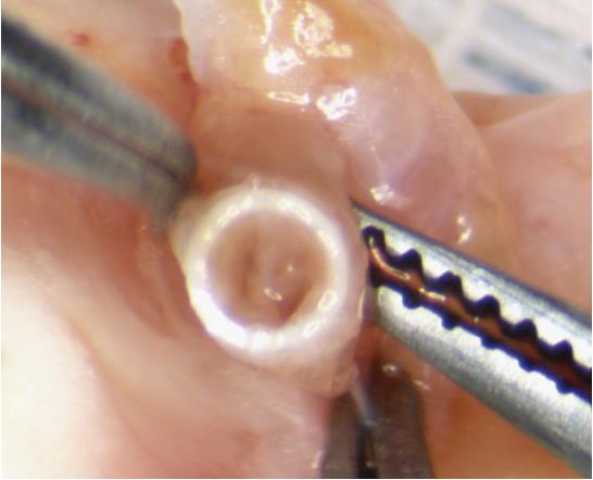
Figure 4 The pulmonary vein forms from the confluence of superior and inferior segmental veins.
The ostia of these veins can be used as landmarks to avoid twisting the everted vein inside the anastomotic cuff.
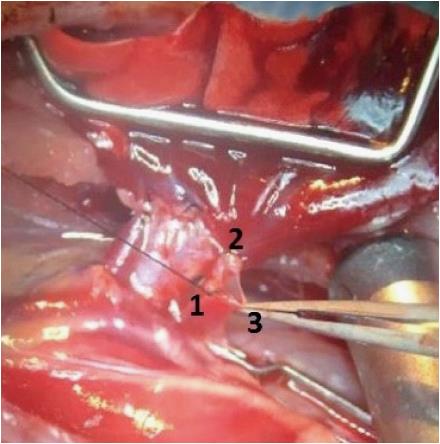
Figure 5 Triangulation of the venotomy by the superior segmental vein ligature (1), pulmonary vein back wall (2) and the tip of the flap (3) results in a wide-mouthed venotomy and serves as a safeguard that allows easy insertion of the donor cuff without tearing the recipient vessel.
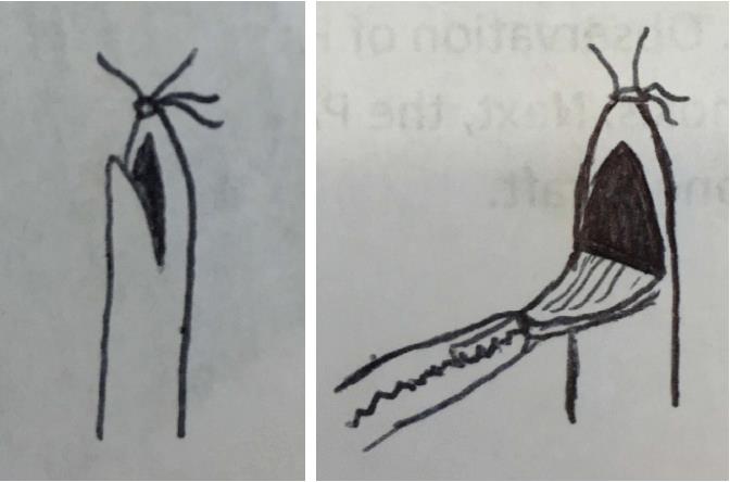
Figure 6 Oblique incision creates a V-shaped flap.
Retraction on this flap with forceps results in a wide mouthed arteritomy that allows easy insertion of the donor cuff.
- Citation: Rajab TK. Anastomotic techniques for rat lung transplantation. World J Transplant 2018; 8(2): 38-43
- URL: https://www.wjgnet.com/2220-3230/full/v8/i2/38.htm
- DOI: https://dx.doi.org/10.5500/wjt.v8.i2.38


