Copyright
©The Author(s) 2017.
World J Transplant. Dec 24, 2017; 7(6): 329-338
Published online Dec 24, 2017. doi: 10.5500/wjt.v7.i6.329
Published online Dec 24, 2017. doi: 10.5500/wjt.v7.i6.329
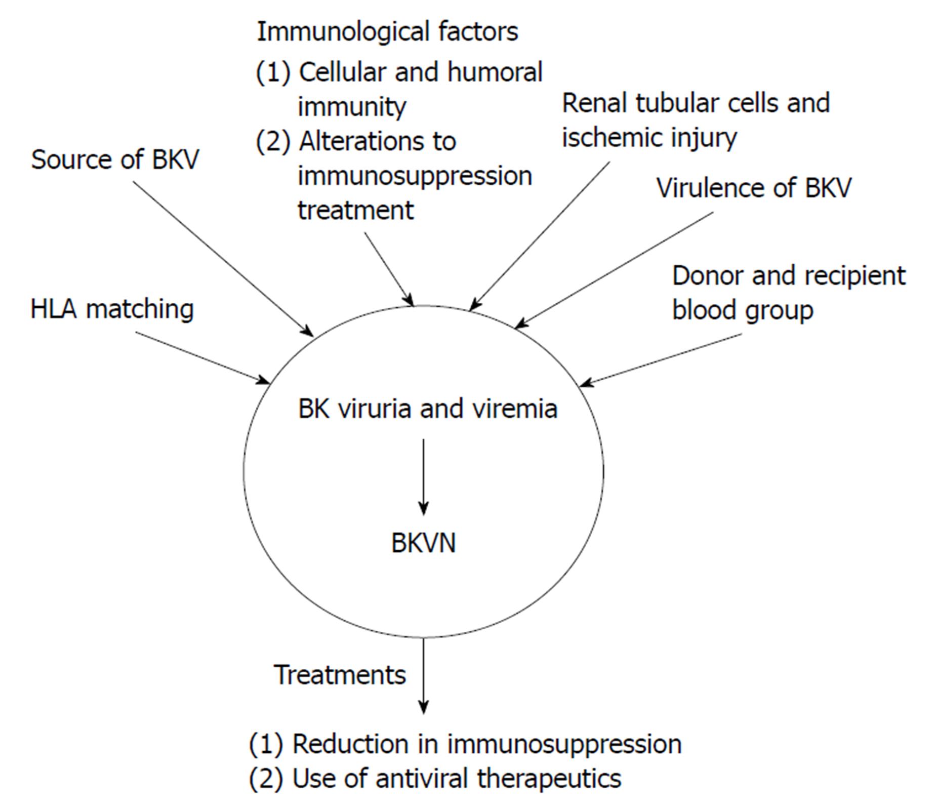
Figure 1 Proposed mechanisms for the pathogenesis of BK virus-associated nephritis after BK virus infection has occurred resulting in BK viruria or BK viremia.
These mechanisms include immunological factors, such as alterations to immunosuppressive therapy and cellular and humoral immunity, the source of BKV, either from the recipient or the donor, HLA matching, donor and recipient blood group. The two main treatment options for BKVN are a reduction in immunosuppression and the use of antiviral therapies. These treatments can also be used for BK viruria and viremia in order to prevent progression to BKVAN. BKV: BK virus; BKVAN: BK virus-associated nephritis.
Figure 2 Histological features of BK virus nephropathy by light microscopy.
A: Tubule-interstitial infiltrate and tubulitis classical for BKVN, but also compatible with any other form of interstitial nephritis such as acute cellular rejection; B: Higher power view of same biopsy sample, with characteristic viral inclusions seen within epithelial cells (circled); C: Positive SV40 immunoperoxidase staining on same specimen, confirming diagnosis of BKVN. BKVN: BK virus nephropathy.
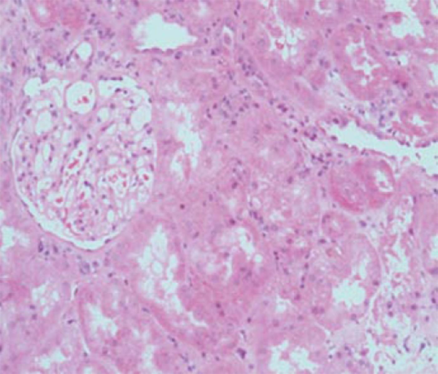
Figure 3 Kidney with preserved tubular architecture, without significant chronic damage or interstitial inflammation, but with BK virus nephropathy confirmed by virtue of positive SV40 staining (as shown in insert taken from immunoperoxidase sample from same biopsy specimen).
Figure 4 Histological features of BK virus nephropathy by electron microscopy.
A: Electron microscopy evidence of viral inclusions (arrow) within epithelial cells, equivalent to those seen and circled in the light microscopy sample shown in Figure 2B; B: Higher power magnification of epithelial viral inclusions; C: Highest magnification demonstrating characteristic appearance and size (labelled) of BK virions.
- Citation: Scadden JR, Sharif A, Skordilis K, Borrows R. Polyoma virus nephropathy in kidney transplantation. World J Transplant 2017; 7(6): 329-338
- URL: https://www.wjgnet.com/2220-3230/full/v7/i6/329.htm
- DOI: https://dx.doi.org/10.5500/wjt.v7.i6.329


