Copyright
©The Author(s) 2017.
World J Transplant. Dec 24, 2017; 7(6): 285-300
Published online Dec 24, 2017. doi: 10.5500/wjt.v7.i6.285
Published online Dec 24, 2017. doi: 10.5500/wjt.v7.i6.285
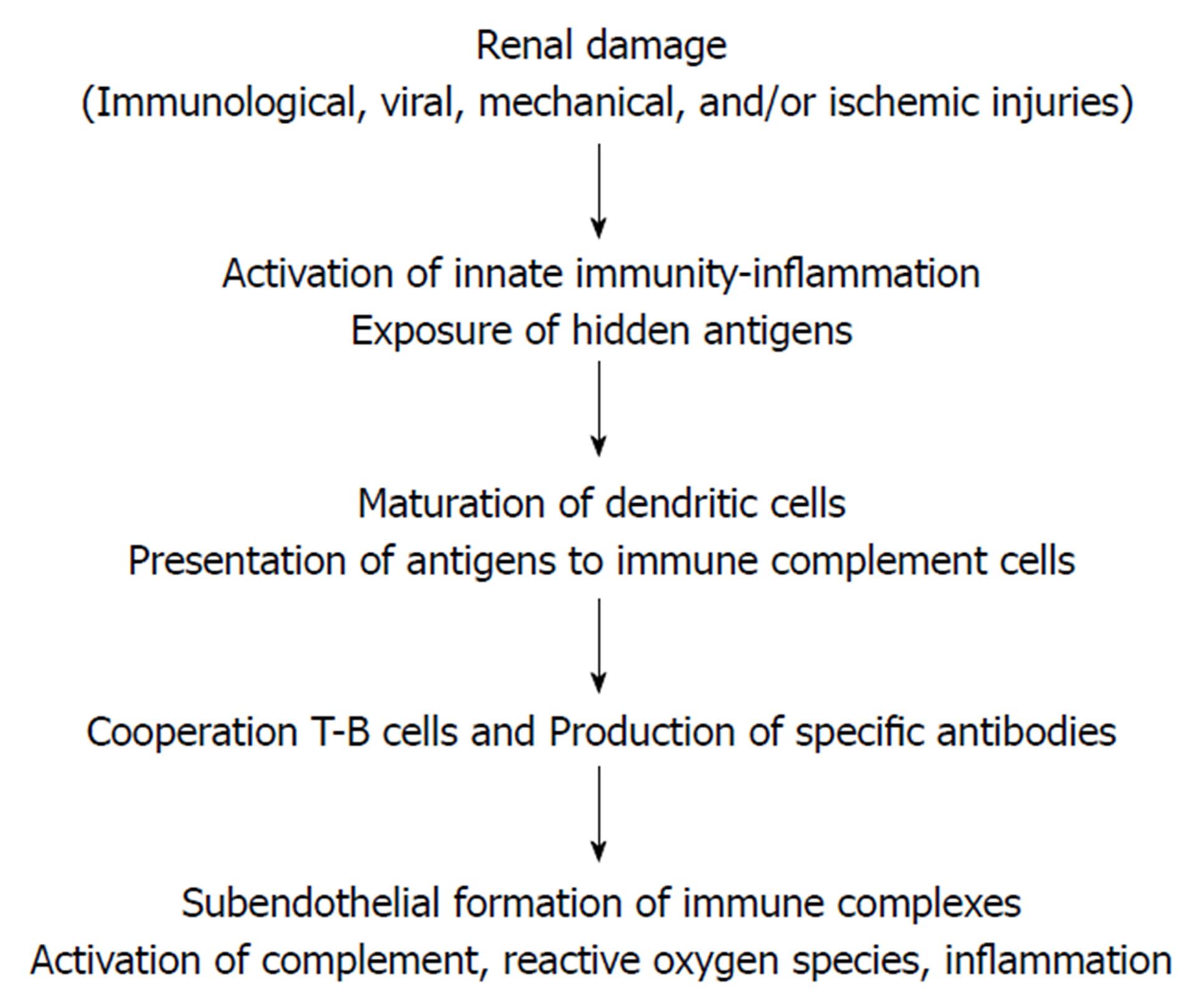
Figure 1 Any type of kidney injury can cause tissue damage.
The danger signals released by the damaged tissue alert the recognition receptors, which activate the inflammatory cells and mediators of the innate immunity system. In this inflammatory environment, hidden podocyte antigens may be exposed, whereas dendritic cells become mature, migrate to lymphatic system, and present the antigen to immune competent cells. T cells cooperate with B cells favoring the production of antibodies directed against the exposed antigens planted in the subepithelium, with in situ formation of immune complexes, activation of complement, formation of free oxygen radicals, and inflammation. Adapted from: Ponticelli et al[10], 2012. De novo membranous nephropathy (MN) in kidney allografts. A peculiar form of all immune disease? With permission.
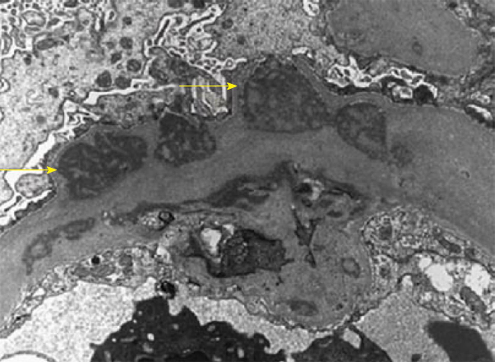
Figure 2 Glomerular capillaries are greatly distorted and thickened by the presence of numerous, sometimes large and/or confluent subendothelial electron-dense deposits (arrows).
The electron-dense deposits have a variegated (“two-toned”) appearance and are finely granular, but they do not show organized substructures. Adapted from: Al-Rabadi et al[73] (2015) (open access).
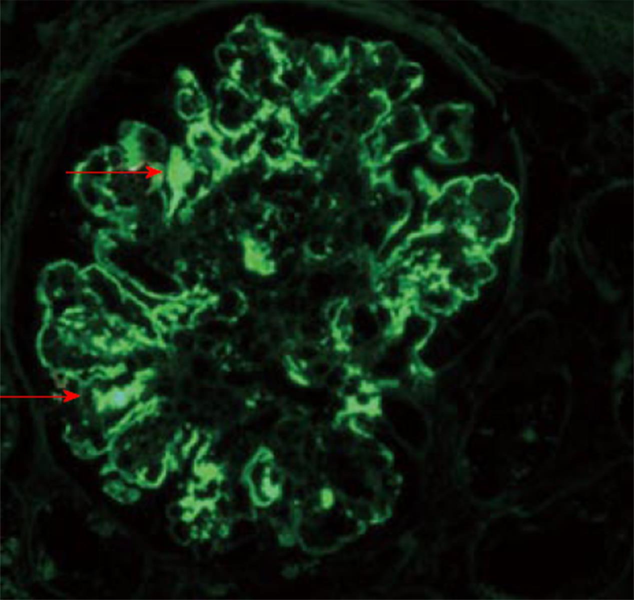
Figure 3 Diffuse irregular granular and pseudo linear deposition of IgG (3+/4+) (arrows).
No staining is found in Bowman’s capsule or the tubular BM. Adapted from: Al-Rabadi et al[73] (2015) (open access).
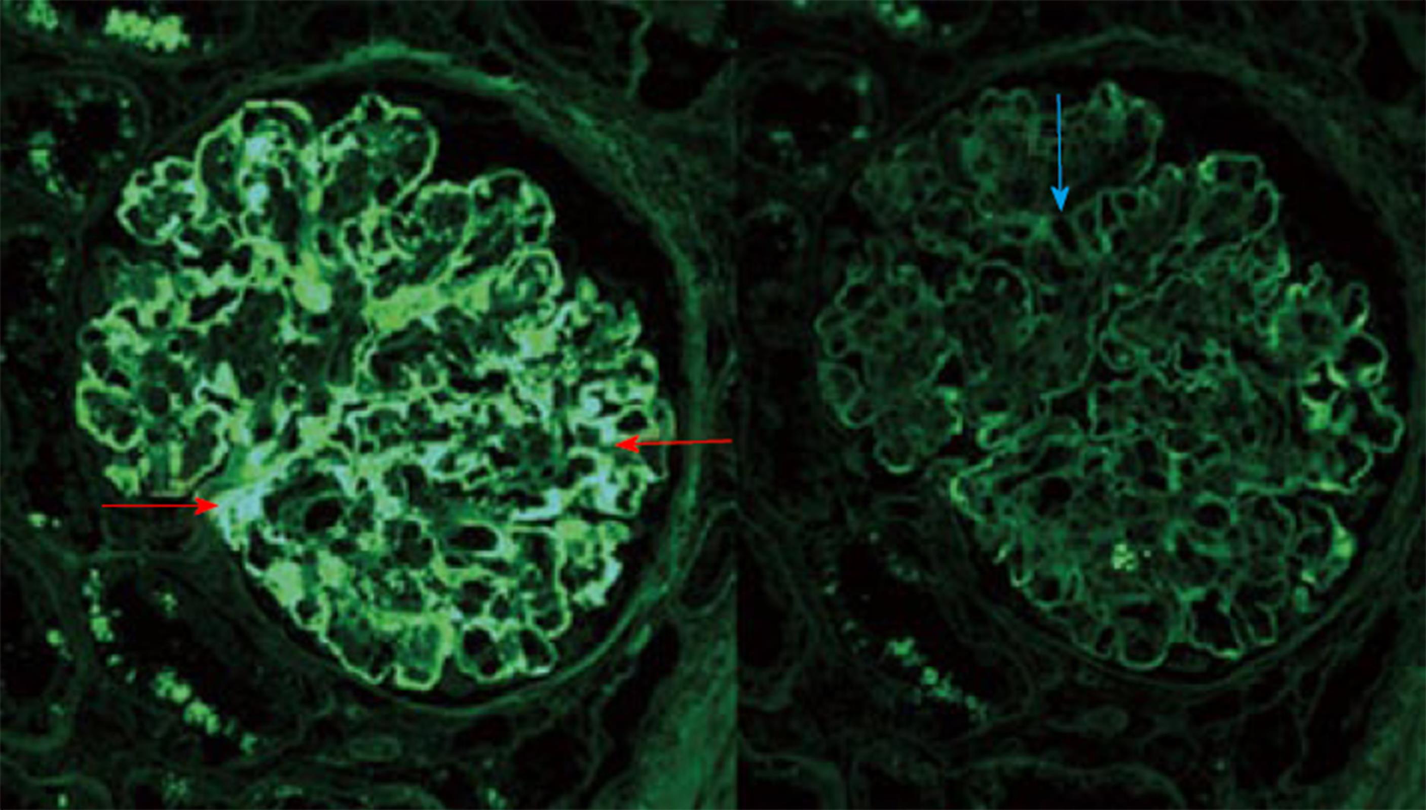
Figure 4 Kappa light chains stain strongly positive (3+/4+) (Rt side, red arrows) along the peripheral capillary walls and mesangial areas.
Lambda light chains are negative in the deposits (Lt side, blue arrow). Adapted from: Al-Rabadi et al[73] (2015) (open access).
- Citation: Abbas F, El Kossi M, Jin JK, Sharma A, Halawa A. De novo glomerular diseases after renal transplantation: How is it different from recurrent glomerular diseases? World J Transplant 2017; 7(6): 285-300
- URL: https://www.wjgnet.com/2220-3230/full/v7/i6/285.htm
- DOI: https://dx.doi.org/10.5500/wjt.v7.i6.285


