Copyright
©The Author(s) 2016.
World J Transplant. Dec 24, 2016; 6(4): 751-758
Published online Dec 24, 2016. doi: 10.5500/wjt.v6.i4.751
Published online Dec 24, 2016. doi: 10.5500/wjt.v6.i4.751
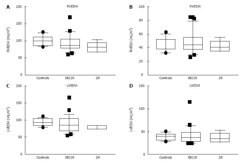
Figure 1 Box and whiskers plots for end-diastolic and end-systolic ventricular volumes of controls and transplant patients without (0R/1R) and with (2R) significant rejection.
A: Right ventricular end-diastolic volume indexed to body surface area (RVEDVi); B: Right ventricular end-systolic volume indexed to body surface area (RVESVi); C: Left ventricular end-diastolic volume indexed to body surface area (LVEDVi); D: Left ventricular end-systolic volume indexed to body surface area (LVESVi).
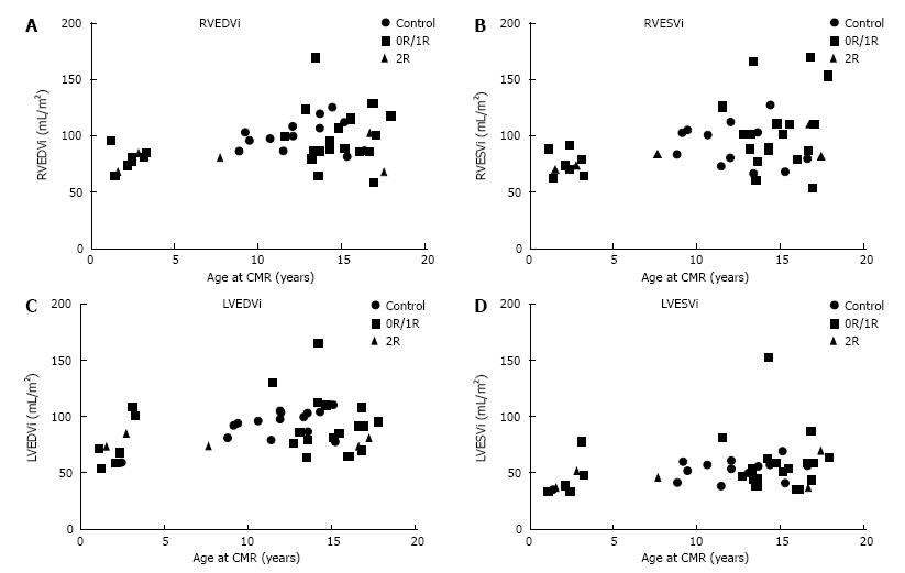
Figure 2 Ventricular volumes plotted as a function of age for controls and transplant patients without (0R/1R) and with (2R) significant rejection.
A: Right ventricular end-diastolic volume indexed to body surface area (RVEDVi); B: Right ventricular end-systolic volume indexed to body surface area (RVESVi); C: Left ventricular end-diastolic volume indexed to body surface area (LVEDVi); D: Left ventricular end-systolic volume indexed to body surface area (LVESVi).
- Citation: Greenway SC, Dallaire F, Kantor PF, Dipchand AI, Chaturvedi RR, Warade M, Riesenkampff E, Yoo SJ, Grosse-Wortmann L. Magnetic resonance imaging of the transplanted pediatric heart as a potential predictor of rejection. World J Transplant 2016; 6(4): 751-758
- URL: https://www.wjgnet.com/2220-3230/full/v6/i4/751.htm
- DOI: https://dx.doi.org/10.5500/wjt.v6.i4.751


