Copyright
©The Author(s) 2016.
World J Transplant. Sep 24, 2016; 6(3): 573-582
Published online Sep 24, 2016. doi: 10.5500/wjt.v6.i3.573
Published online Sep 24, 2016. doi: 10.5500/wjt.v6.i3.573
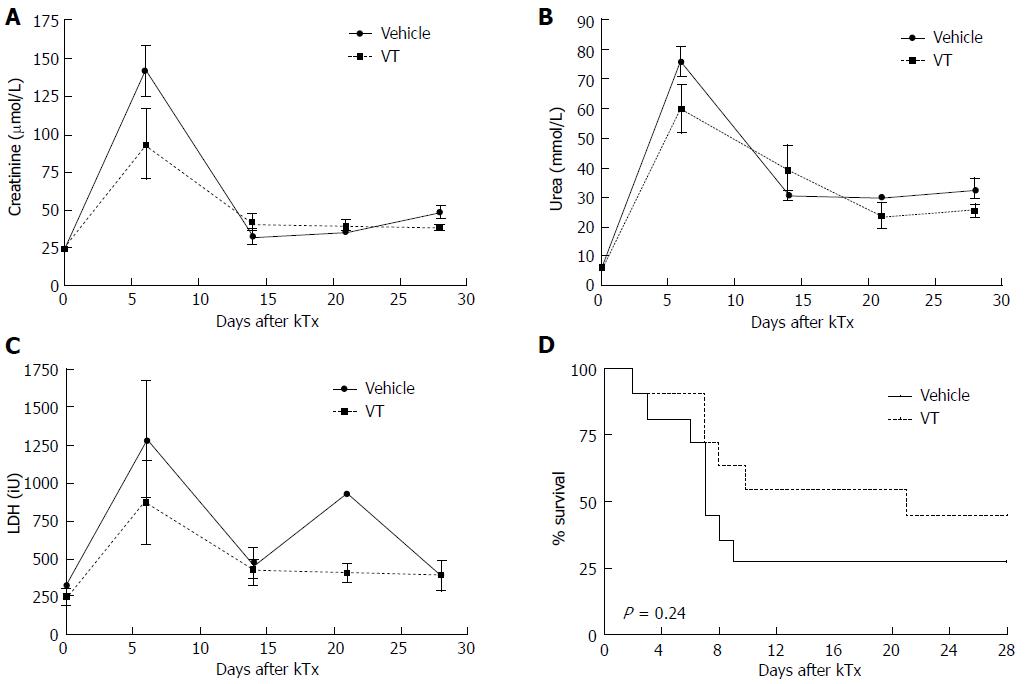
Figure 1 Vasculotide shows trends to improved kidney function and survival in an MHC-mismatched renal transplant model.
C57Bl/6 donor mice received 500 ng VT or vehicle prior to surgery (-1 h). Balb/c recipients were injected with 500 ng VT i.p. or vehicle directly and on day 3 after kidney transplantation. A-C: Kidney function [serum creatinine, urea and lactate dehydrogenase (LDH) levels] was monitored on day 6, 14, 21 and 28 after transplantation in murine serum and analyzed with unpaired t test (day 6, n = 7-10); D: Kaplan-Meier survival after MHC-mismatch kidney transplantation (n = 11 per group). VT: Vasculotide.
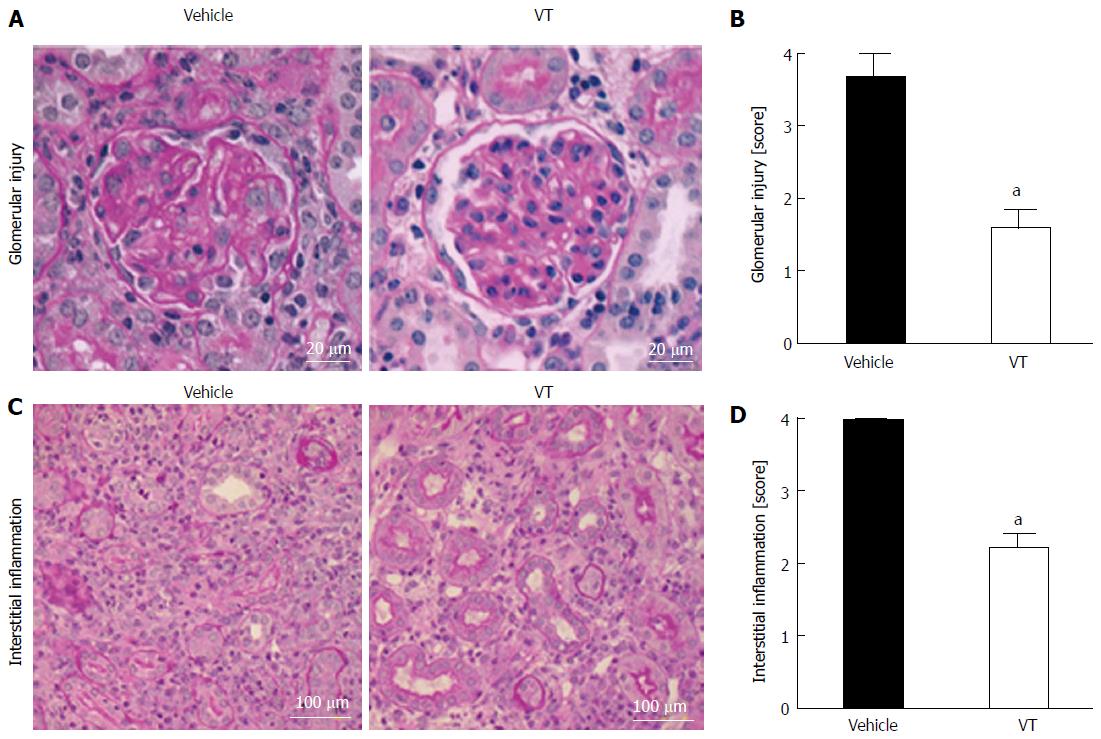
Figure 2 Vasculotide-treated mice show less infiltration of inflammatory cells in the kidney.
A: Exemplary PAS staining of kidneys from mice treated with vehicle or VT. Glomerular injury was evaluated on day 28 after transplantation; B: Semi-quantification of glomerular injury by surveying kidney cross-sections. Scoring was done as described above (see Table 1) (vehicle n = 3; VT n = 5) (aP < 0.05); C: PAS staining for peritubular injury in transplanted kidneys from mice treated with vehicle or VT; D: Semi-quantification of interstitial inflammation by surveying kidney cross-sections from transplanted mice (aP < 0.05). Scoring was done as described above (Table 1) (vehicle n = 3; VT n = 5). Semi-quantification was assessed using Mann-Whitney test. VT: Vasculotide; PAS: Periodic acid Schiff.
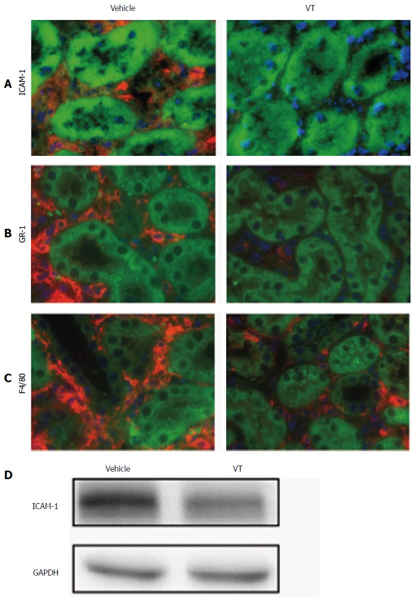
Figure 3 Vasculotide treatment reduces vascular inflammation and tissue infiltration in kidney transplantation.
Fluorescent immunohistochemistry for (A) ICAM-1 (red), (B) Gr-1 (red) and (C) F4/80 (red) in kidney cross-sections of transplanted mice (vehicle or VT-treated) on day 28 after transplantation. Autofluorescence is shown in green. Images are exemplary for n = 5/condition; D: Immunoblot of murine kidney homogenate for ICAM-1 and GAPDH for the same conditions. VT: Vasculotide; ICAM-1: Intercellular adhesion molecule; GAPDH: Glyceraldehyde 3-phosphate dehydrogenase.
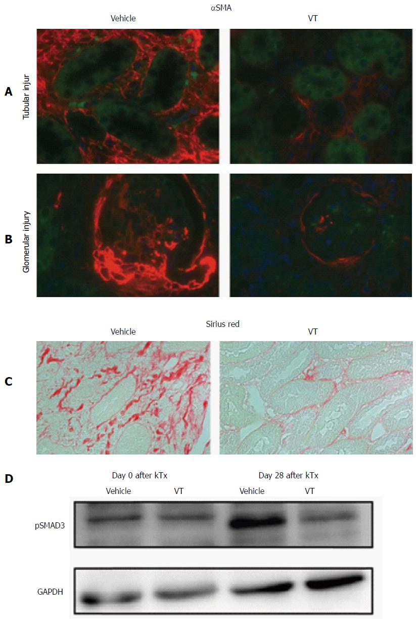
Figure 4 Vasculotide ameliorates fibrogenesis.
Fluorescent immunohistochemistry of α-smooth-muscle-actin (red) regarding (A) tubular and (B) glomerular injury in kidney cross-sections of transplanted mice (vehicle or VT-treated) on day 28 after transplantation. Autofluorescence is shown in green. Images are exemplary for n = 5/condition; C: Sirius red staining for the same conditions (D) Immunoblot of murine kidney homogenates for pSMAD3 in healthy (day 0) and kidney transplanted mice (day 28) upon vehicle or VT treatment. VT: Vasculotide; αSMA: α-smooth-muscle-actin; GAPDH: Glyceraldehyde 3-phosphate dehydrogenase.
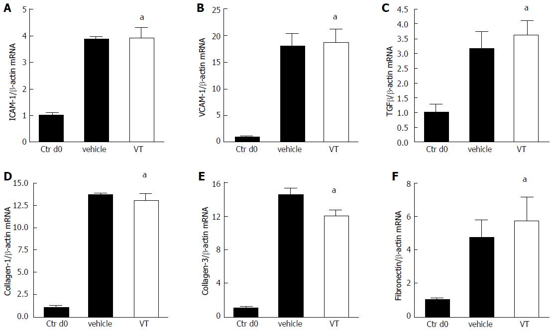
Figure 5 Anti-inflammatory properties of vasculotide are not regulated on the transcriptional level.
A-F: Mice were treated with vehicle (n = 3) or VT (n = 5) and underwent kidney transplantation. Transplanted kidneys were harvested on day 28 after transplantation. Explanted donor kidneys from day 0 served as control (n = 3). Expression levels of Intercellular adhesion molecule (ICAM-1), Vascular cell adhesion protein 1 (VCAM-1), Transforming growth factor β (TGFβ), collagen-1, collagen-3 and fibronectin in kidney homogenates were determined via RT-qPCR and analyzed using Mann-Whitney test (aP < 0.05). VT: Vasculotide.
- Citation: Thamm K, Njau F, Van Slyke P, Dumont DJ, Park JK, Haller H, David S. Pharmacological Tie2 activation in kidney transplantation. World J Transplant 2016; 6(3): 573-582
- URL: https://www.wjgnet.com/2220-3230/full/v6/i3/573.htm
- DOI: https://dx.doi.org/10.5500/wjt.v6.i3.573


