Copyright
©The Author(s) 2017.
World J Clin Infect Dis. Aug 25, 2017; 7(3): 46-49
Published online Aug 25, 2017. doi: 10.5495/wjcid.v7.i3.46
Published online Aug 25, 2017. doi: 10.5495/wjcid.v7.i3.46
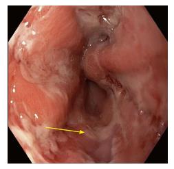
Figure 1 Diffuse ulcerations in the mid esophagus with inflammatory exudate, severe esophagitis 32-42 cm in esophagus.
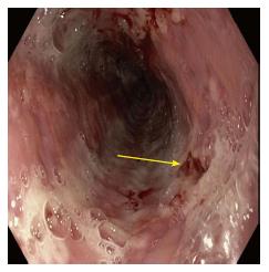
Figure 2 Endoscopic appearance of shallow base ulcers with evidence of erosions and superficial bleeding of the esophagus.
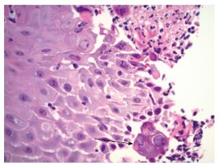
Figure 3 Hematoxylin and Eosin stain showing squamous cells with viral cytopathic effects of herpes simplex virus 1 and 2 - cowdry type A inclusion body- which includes multinucleation, margination of chromatin and molding of nuclei.
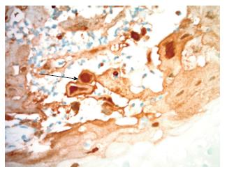
Figure 4 Immunohistochemistry stain for herpes simplex virus 1 and 2 shows the involved nuclei as staining brown for herpes simplex virus viral particles.
- Citation: de Choudens FCR, Sethi S, Pandya S, Nanjappa S, Greene JN. Atypical manifestation of herpes esophagitis in an immunocompetent patient: Case report and literature review. World J Clin Infect Dis 2017; 7(3): 46-49
- URL: https://www.wjgnet.com/2220-3176/full/v7/i3/46.htm
- DOI: https://dx.doi.org/10.5495/wjcid.v7.i3.46


