Copyright
©2012 Baishideng Publishing Group Co.
World J Clin Infect Dis. Aug 25, 2012; 2(4): 63-76
Published online Aug 25, 2012. doi: 10.5495/wjcid.v2.i4.63
Published online Aug 25, 2012. doi: 10.5495/wjcid.v2.i4.63
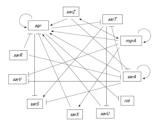
Figure 1 Proposed genetic regulatory network involving sarA family genes and agr in Staphylococcus aureus.
The model is constructed from published studies[25,29,31,86,101,105,106,109,110,114,115,120-125,129,130,135,141,144] that are mostly based on a limited number of laboratory strains. Therefore, it may not be entirely applicable to all strains. Arrows indicate activation; blocked arrows indicate repression.
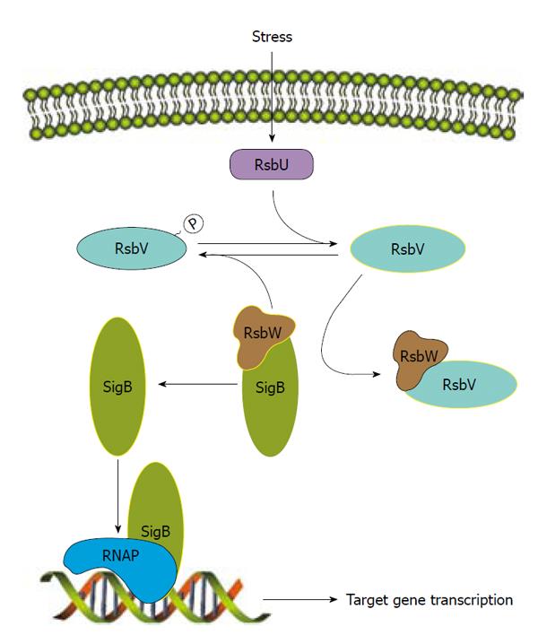
Figure 2 Post-transcriptional regulation of SigB.
After stress-induction, RsbU de-phosphorylates RsbV, which can then bind specifically to RsbW thereby removing RsbW from SigB. Phosphorylated RsbV is inactive and therefore cannot bind RsbW. RsbW also promotes phosphorylation of RsbV to maintain its inactivity. RsbW binds to SigB to inhibit transcription by preventing SigB from complexing with the RNA polymerase (RNAP). Once SigB is free from inhibition by RsbW, it can complex with RNAP forming the holoenzyme and activate transcription of target genes. Active proteins are highlighted with yellow.
-
Citation: Junecko JM, Zielinska AK, Mrak LN, Ryan DC, Graham JW, Smeltzer MS, Lee CY. Transcribing virulence in
Staphylococcus aureus . World J Clin Infect Dis 2012; 2(4): 63-76 - URL: https://www.wjgnet.com/2220-3176/full/v2/i4/63.htm
- DOI: https://dx.doi.org/10.5495/wjcid.v2.i4.63


