Copyright
©The Author(s) 2020.
World J Exp Med. Apr 27, 2020; 10(3): 26-40
Published online Apr 27, 2020. doi: 10.5493/wjem.v10.i3.26
Published online Apr 27, 2020. doi: 10.5493/wjem.v10.i3.26
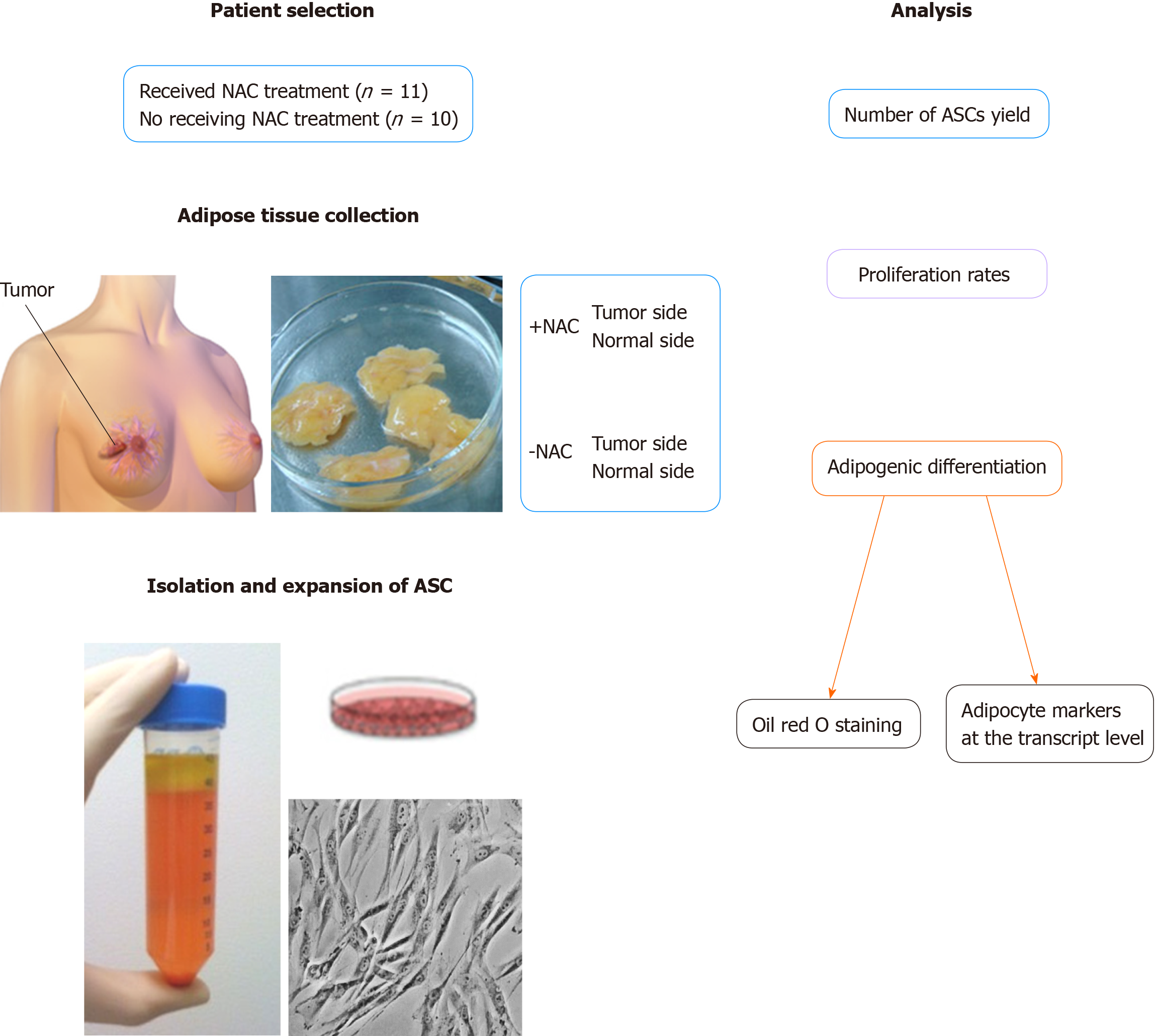
Figure 1 Experimental design.
Adipose-derived stromal cells (ASCs) were isolated from both the cancerous side and noncancerous side of the breast from the same patients with receiving neoadjuvant chemotherapy (NAC) treatment or not-receiving NAC. The number of ASCs yield, proliferation rates and the ability to undergo adipogenic differentiation was analyzed by constructing growth curves, quantitative RT-PCR and histological staining and compared to the ASCs obtained from patients not-receiving NAC treatment. ASCs: Adipose-derived stromal cells; NAC: Neoadjuvant chemotherapy.
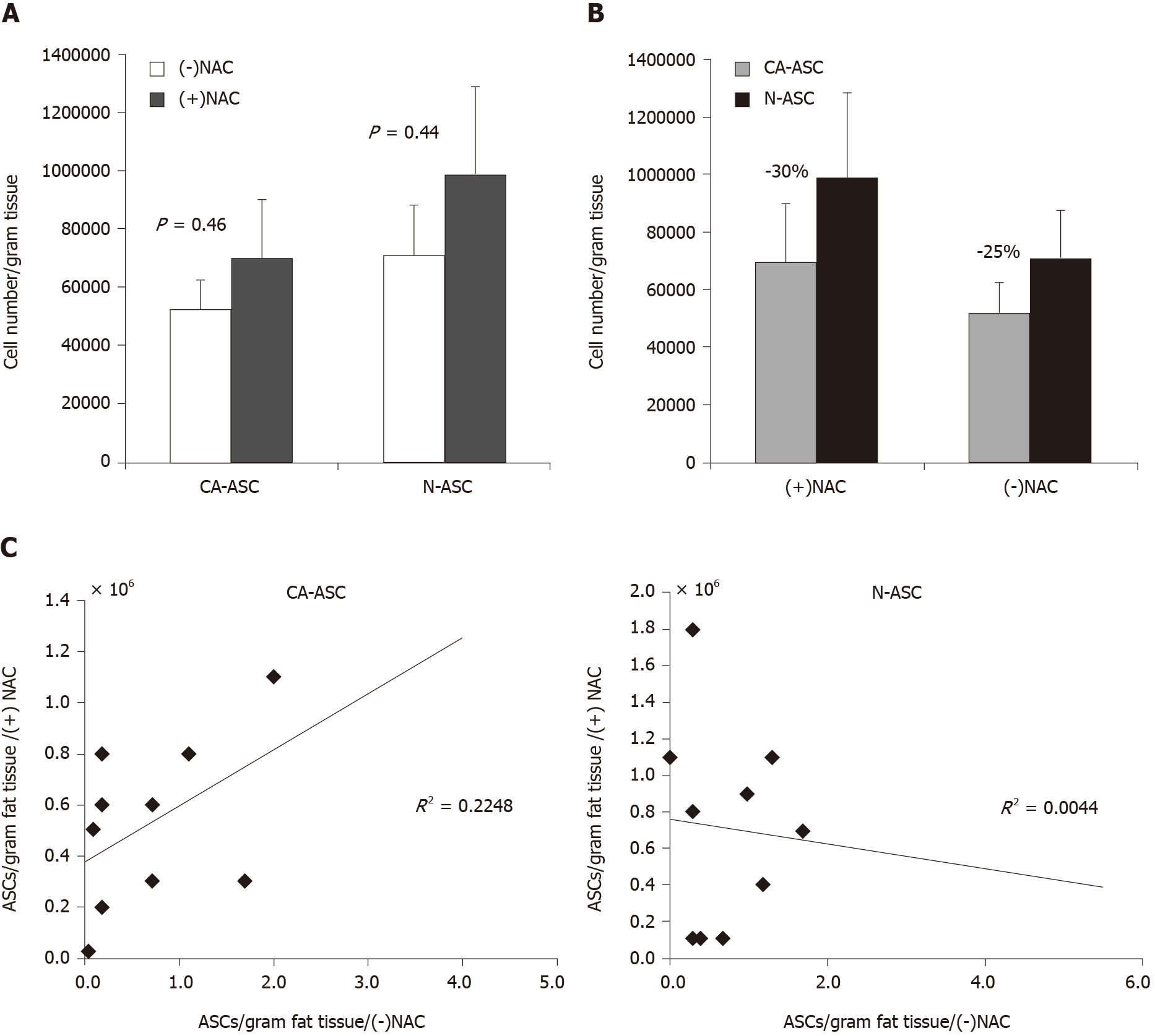
Figure 2 Isolation of adipose-derived stromal cells from breast tissues in patients after received neoadjuvant chemotherapy treatment.
A: Number of ASCs isolated per gram of adipose tissue from the cancerous side (CA-ASC) of the breast and noncancerous side (N-ASC) of the breast in patients receiving neoadjuvant chemotherapy (+NAC) vs patients not-receiving neoadjuvant chemotherapy (-NAC). B: ASCs yield from the cancerous side of the breast was lower than the noncancerous side of the breast in both patients receiving NAC and not-receiving NAC. C: Overall correlation between the numbers of harvesting ASCs from the cancerous side and noncancerous side of the breast in patients receiving NAC or not-receiving NAC (n = 21, mean ± SE, P = NS). ASCs: Adipose-derived stromal cells; NAC: Neoadjuvant chemotherapy.
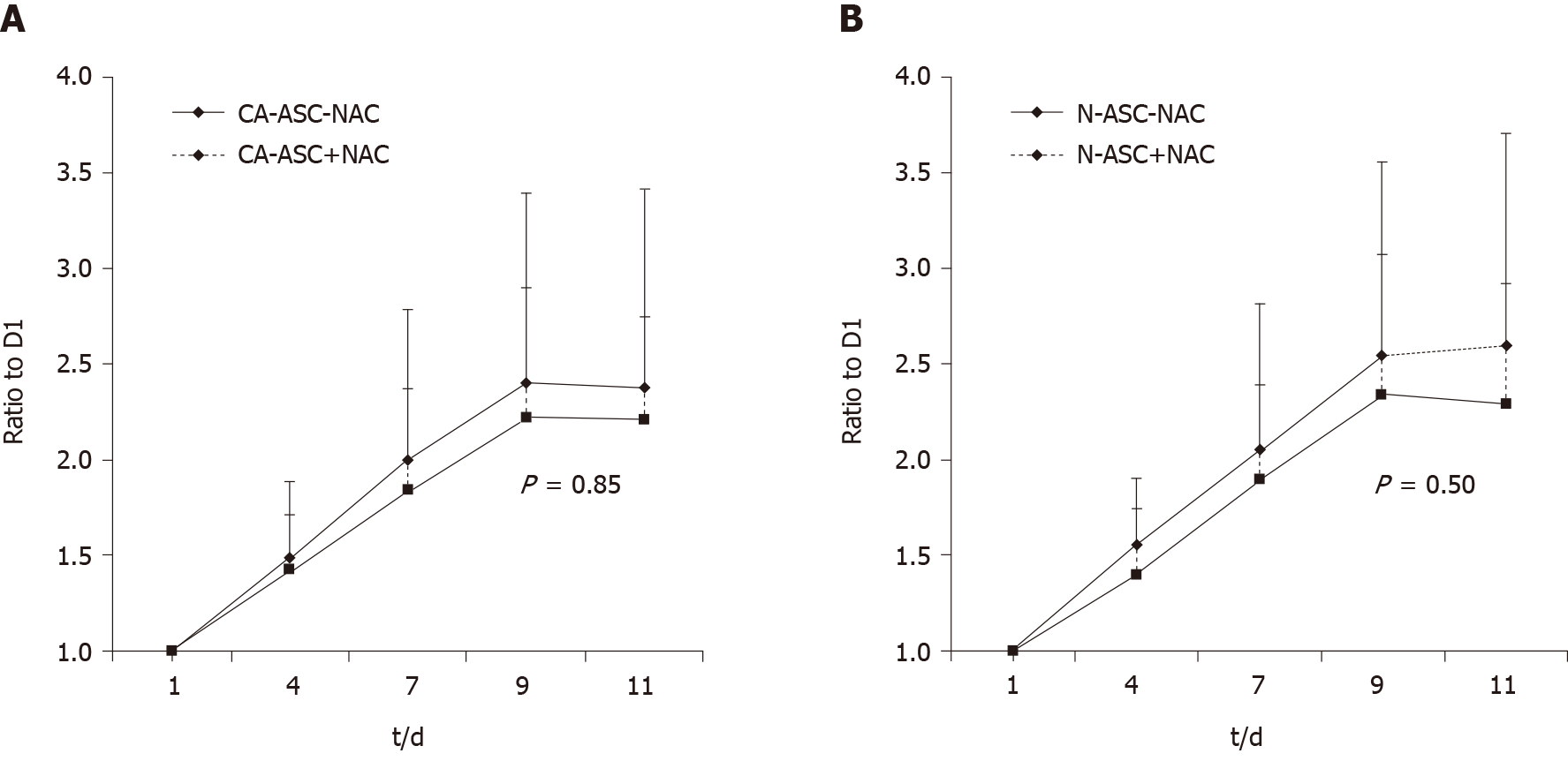
Figure 3 Proliferation of adipose-derived stromal cells in patients after received neoadjuvant chemotherapy treatment.
A: Growth curve of ASCs from the cancerous side (CA-ASC) of the breast in patients receiving neoadjuvant chemotherapy (+NAC) vs not-receiving neoadjuvant chemotherapy (-NAC) during 11 d culturing. B: Comparison of ASCs from the noncancerous side (N-ASC) of the breast vs cancerous side (CA-ASC) of the breast on cell proliferation between patients receiving NAC treatment and not-receiving NAC during 11 d culturing (n = 21, mean ± SE, P = NS). ASC: Adipose-derived stromal cell; NAC: Neoadjuvant chemotherapy.
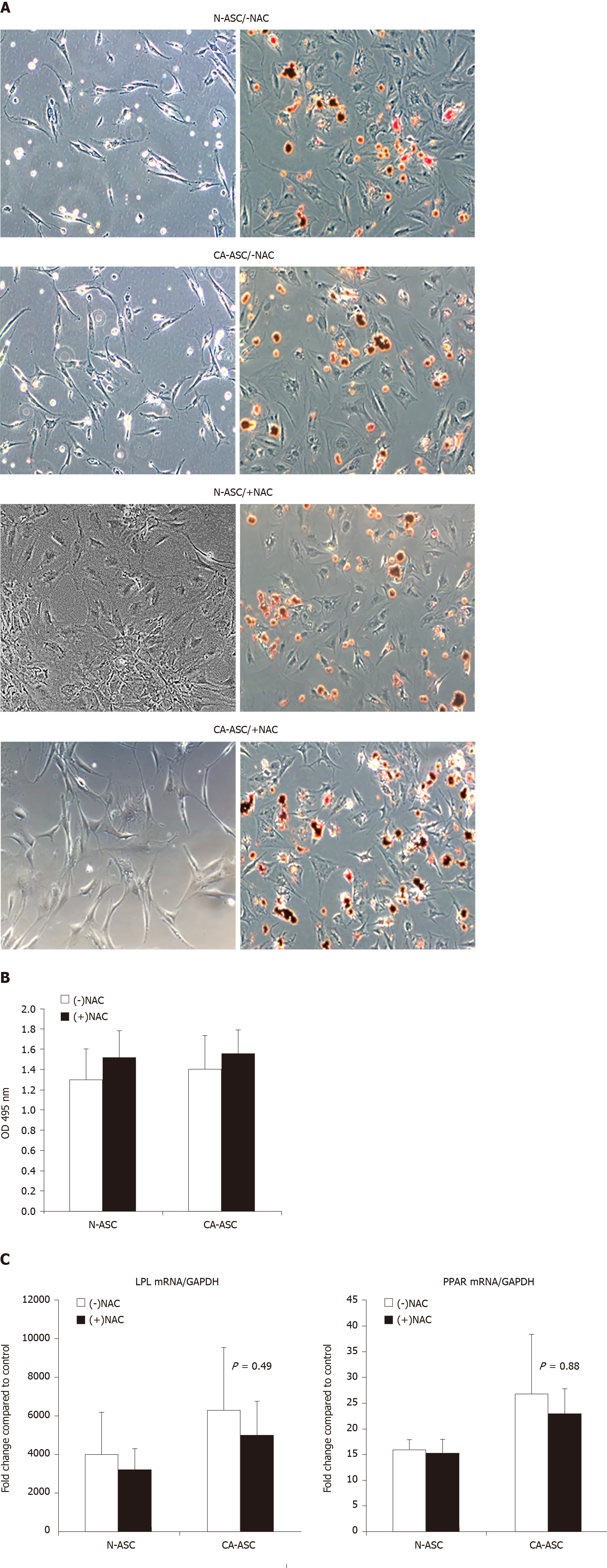
Figure 4 Adipogenic differentiation capacity of adipose-derived stromal cells in patients after received neoadjuvant chemotherapy treatment.
A: Oil red O stains in the adipogenic-induced ASCs from the cancerous side of the breast (CA-ASC) and noncancerous side of the breast (N-ASC) in patients receiving NAC treatment (+NAC) vs not-receiving NAC (-NAC) from undifferentiated ASCs (left) and differentiated ASCs (right). B: Quantified by extracting the dyes for evaluating the adipogenic differentiated cells levels from the cancerous side of the breast or noncancerous side of the breast ASCs in patients receiving NAC treatment vs not-receiving NAC. C: Real-time PCR evaluating the mRNA expression of peroxisome proliferator activated receptor gamma (PPARγ) and lipoprotein lipase (LPL) in differentiating ASCs from the cancerous side of the breast or noncancerous side of the breast in patients receiving NAC treatment vs not-receiving NAC (n = 21, mean ± SE, P = NS). ASC: Adipose-derived stromal cell; NAC: Neoadjuvant chemotherapy.
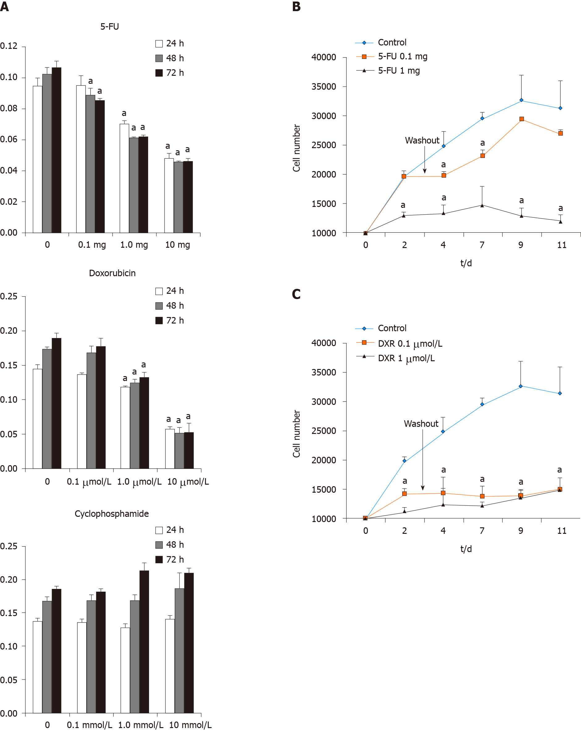
Figure 5 Recovery of adipose-derived stromal cells analysis following exposure to individual chemotherapeutic agents in vitro human population.
A: ASCs were isolated from female patients during reconstructive procedures. Cell viability was measured by MTT assays after ASCs treated with different dosages of 5-FU, DXR and cyclophosphamide on 24, 48, and 72 h. B: Growth curves of ASCs treated with 5-FU or DXR for 72 h then removed and the cells were cultured in medium without drugs for an additional 8 d. The results represent the mean ± SE of triplicate cultures of three representative experiments. n = 3, aP < 0.05 vs control. 5-FU: 5-fluorouracil; DXR: Doxorubicin.
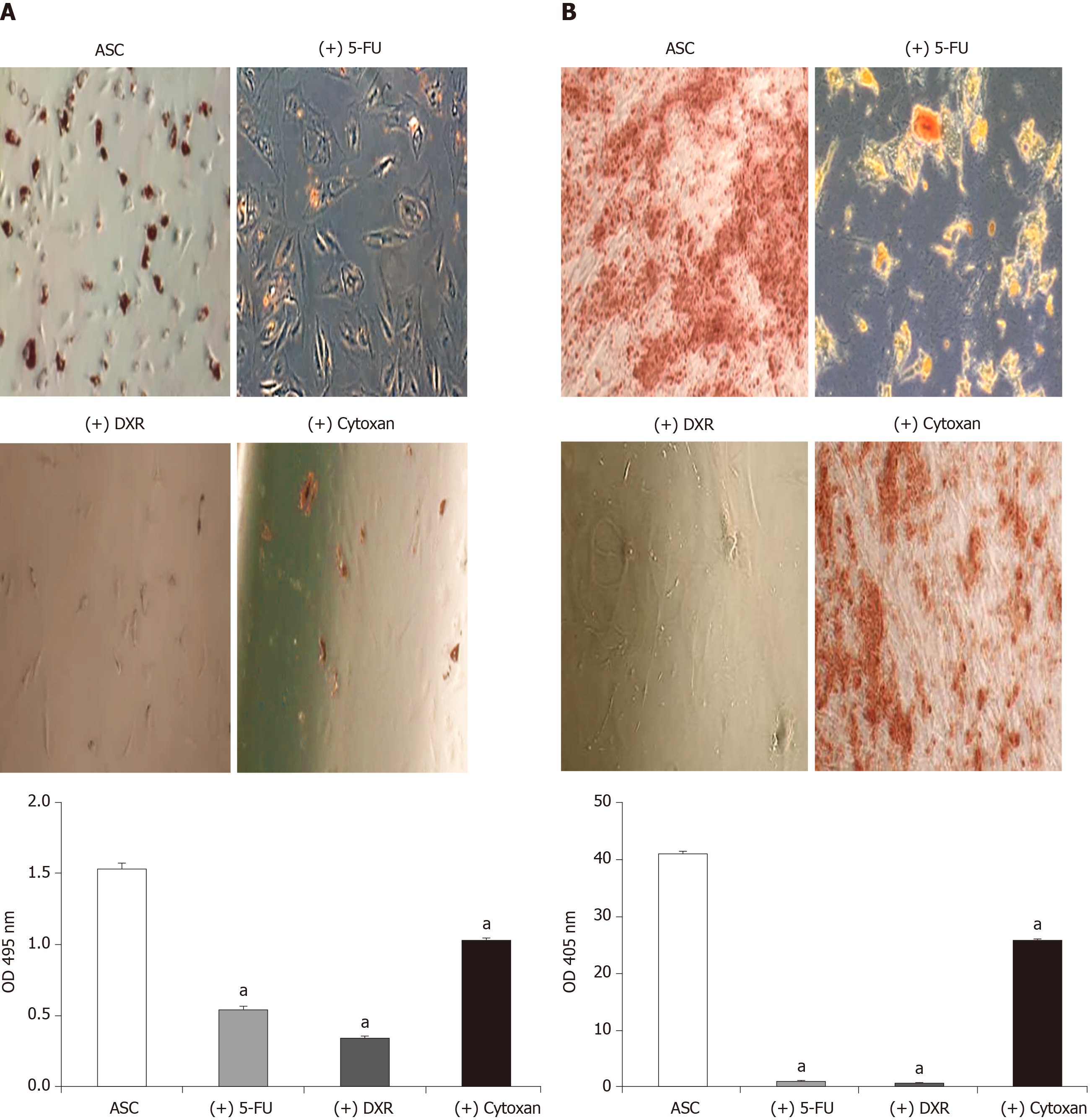
Figure 6 Differentiation capacity of adipose-derived stromal cells analysis following exposure to individual chemotherapeutic agents in vitro human population.
A: Oil red O stains, quantified by extracting the dyes for evaluating the adipogenic differentiated cell levels. B: Alizarin red stains, quantified by extracting the dyes for evaluating the osteogenic differentiated cell levels. ASCs were isolated from female patients during reconstructive procedures. n = 3, mean ± SE, aP < 0.05 vs control. ASC: Adipose-derived stromal cell; 5-FU: 5-fluorouracil; DXR: Doxorubicin; OD: Optical density.
- Citation: Hagaman AR, Zhang P, Koko KR, Nolan RS, Fromer MW, Gaughan J, Matthews M. Isolation and identification of adipose-derived stromal/stem cells from breast cancer patients after exposure neoadjuvant chemotherapy. World J Exp Med 2020; 10(3): 26-40
- URL: https://www.wjgnet.com/2220-315x/full/v10/i3/26.htm
- DOI: https://dx.doi.org/10.5493/wjem.v10.i3.26


