Copyright
©The Author(s) 2019.
World J Crit Care Medl. Sep 11, 2019; 8(5): 82-86
Published online Sep 11, 2019. doi: 10.5492/wjccm.v8.i5.82
Published online Sep 11, 2019. doi: 10.5492/wjccm.v8.i5.82
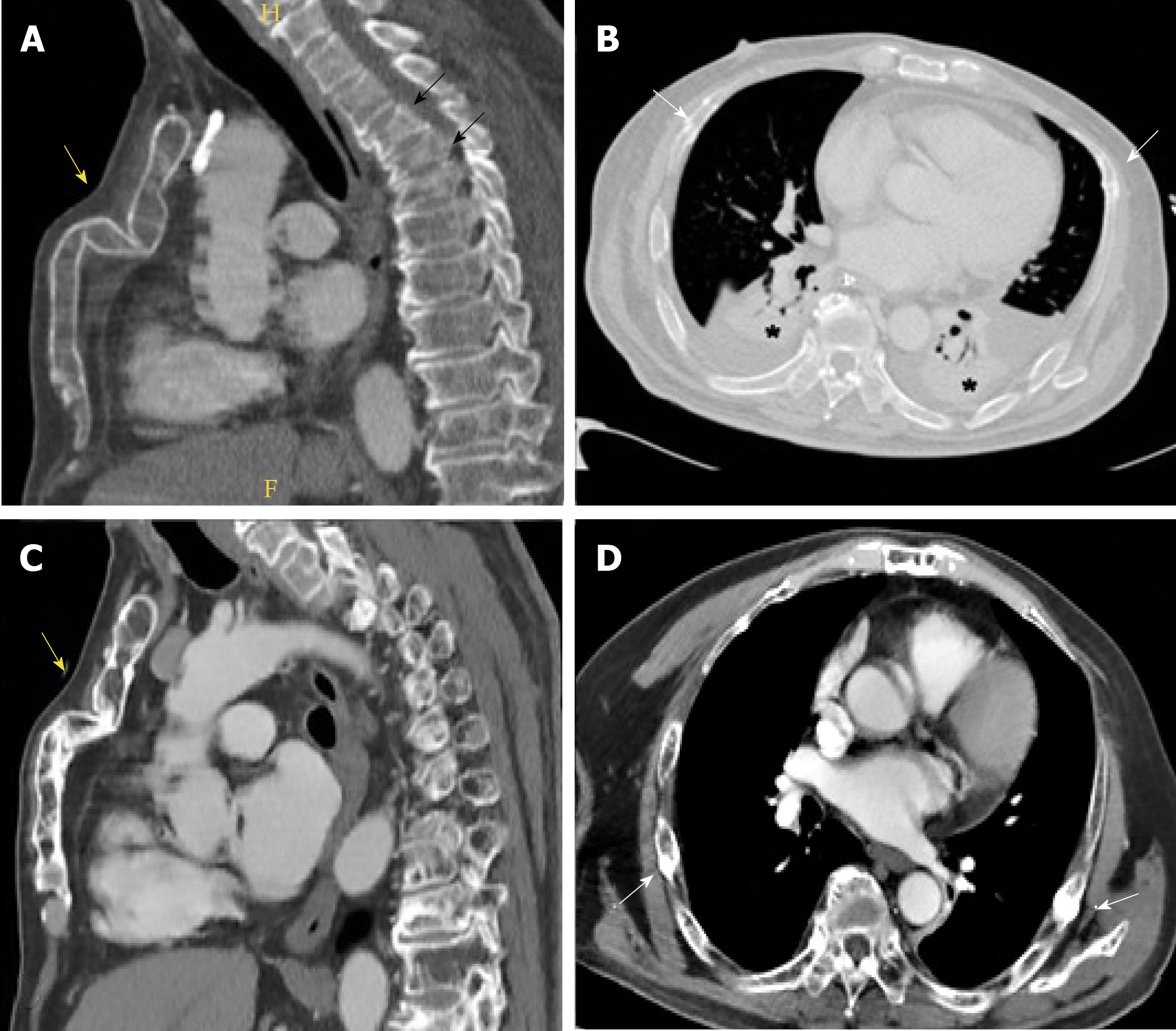
Figure 1 Sagittal computed tomography image.
A: A depressed pathological fracture of the sternum (yellow arrow) resulting in an evident deformity of the anterior chest wall. Multiple spontaneous compression fractures of the thoracic spine can also be observed (black arrows); B: Axial computed tomography (CT) image demonstrates multiple rib fractures (white arrows) associated to bilateral lung consolidations (asterisks) and pleural effusion; C and D: Control CT scan, realized months later, shows important improvement of both sternal (C, yellow arrow) and rib fractures (D, white arrows), with the appearance of fracture calluses. Complete resolution of lung consolidations and pleural effusion (D).
- Citation: Muñoz-Bermúdez R, Abella E, Zuccarino F, Masclans JR, Nolla-Salas J. Successfully non-surgical management of flail chest as first manifestation of multiple myeloma: A case report. World J Crit Care Medl 2019; 8(5): 82-86
- URL: https://www.wjgnet.com/2220-3141/full/v8/i5/82.htm
- DOI: https://dx.doi.org/10.5492/wjccm.v8.i5.82


